

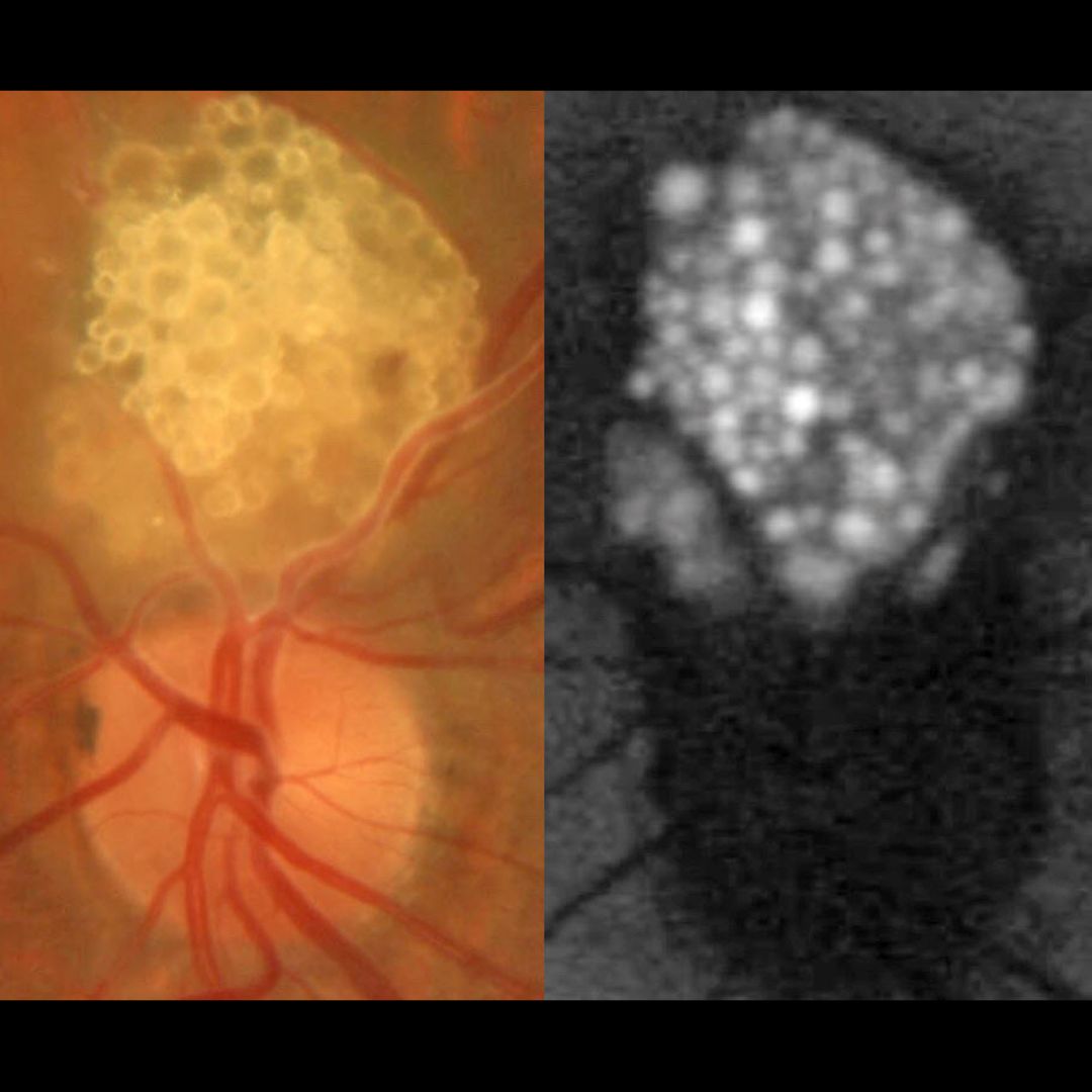
This healthy 43 YO male presented with an asymptomatic lesion in his left eye. Family history was negative. Vision was 20/20 in his normal OD and 20/20 OS.
Color photography shows a glistening yellow-white inner retinal lesion just superior to the optic nerve composed of innumerable semi-transparent spherules which hyper-autofluoresce.
Learning Points:
A retinal astrocytic hamartoma is a benign tumor composed of spindle-shaped fibrous astrocytes located in the nerve fiber layer, most commonly associated with tuberous sclerosis. Tuberous sclerosis includes the classic triad of seizures, mental deficiency and fibroangiomas, but patients can have lesions throughout the body including the eyes, skin (ash-leaf spots), bone, nails, teeth, brain, heart, lungs and kidneys. Early in life the inner retinal lesions have a fleshy white appearance and are often multifocal and bilateral. With time, as in this patient, they show a more classic “mulberry appearance” with significant calcification. Rarely, the vascular component can cause intraretinal exudation requiring treatment.
Clinically retinocytomas can resemble astrocytic hamartomas. However, retinocytomas often have underlying RPE changes and chorioretinal scarring (lacking in our patient), which are absent in astrocytic hamartomas.
Since the patient was self-paying, neurologic imaging was not performed and observation was recommended. A neurology reference was given.



