

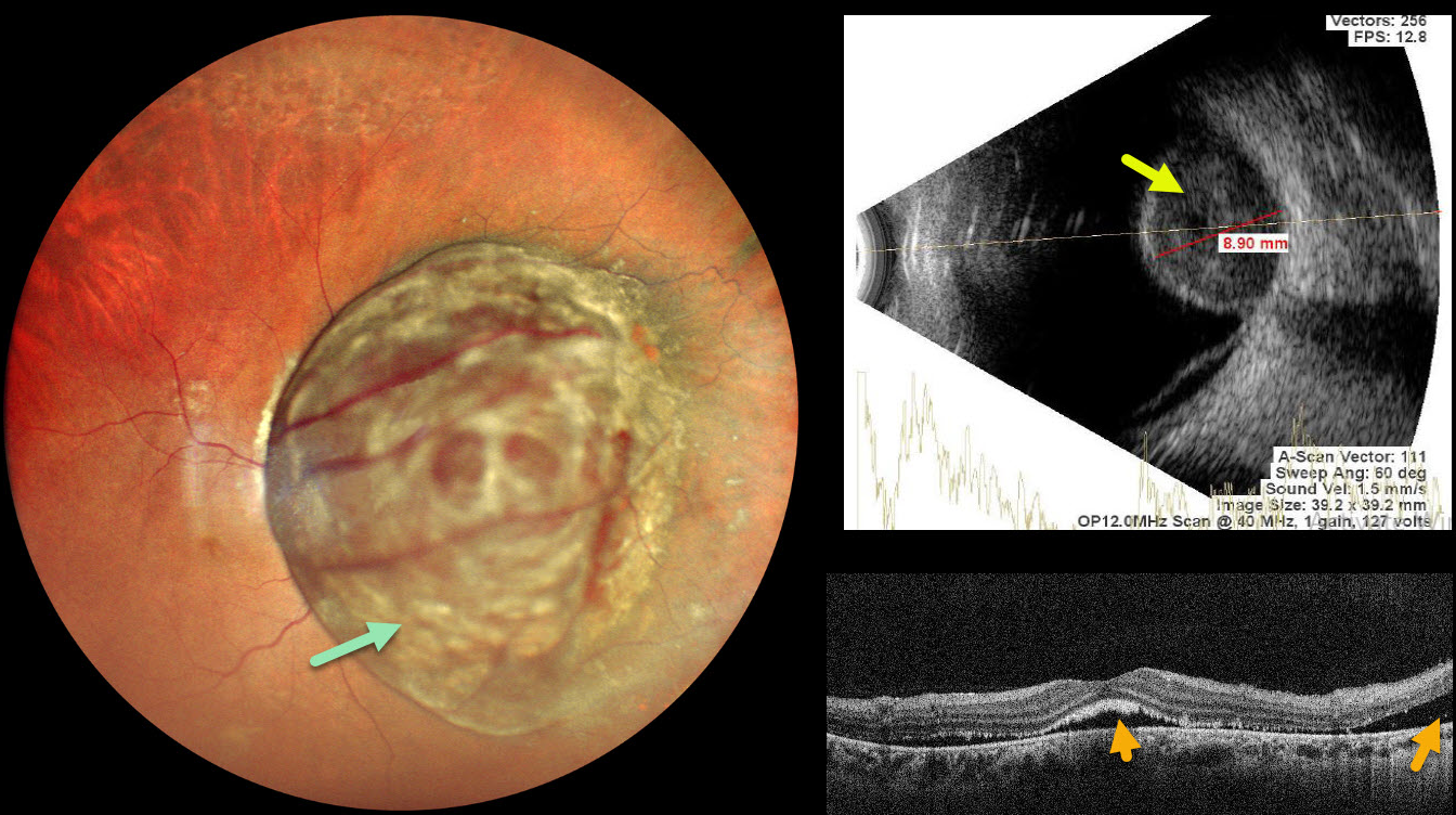
This 47YO female presented with 1 month of decreased vision in her right eye. Vision was 20/400 OD and 20/20 in her normal OS.
Pseudocolor SLO imaging shows a large, well-circumscribed pigmented choroidal lesion that obscures the entire posterior pole. The tumor vessels are markedly dilated and tortuous. OCT shows foveal and nasal subretinal fluid with ragged shed outer segments along the posterior retinal surface. The lesion, which measures 8.9mm in thickness, shows moderate homogenous internal reflectivity on B-scan ultrasonography. She was referred to ocular oncology for further evaluation and treatment.
Learning Points:
Uveal melanoma is a malignant tumor arising from melanocytes in the uveal tract (iris, ciliary body, or choroid). The choroid is the most common site, accounting for 90% of uveal melanomas. Uveal melanoma is the most common primary intraocular cancer in adults, whereas metastatic disease is the most common intraocular malignancy. The liver is the most common metastatic site, although metastatic disease is rare on presentation. Metastases are more common with large tumors, BAP1 mutations, and certain tumor-related genetic markers (including positive PRAME and Class 2).



