

This 4YO girl’s parents noticed that her right eye was deviating outwards for several months, along with decreased vision bilaterally. Her mother gave a history of prematurity at 28 weeks of gestational age with a low birth weight of 1175 g and oxygen exposure. Vision was counting fingers OD and 20/80 OS. There was a large right exotropia.
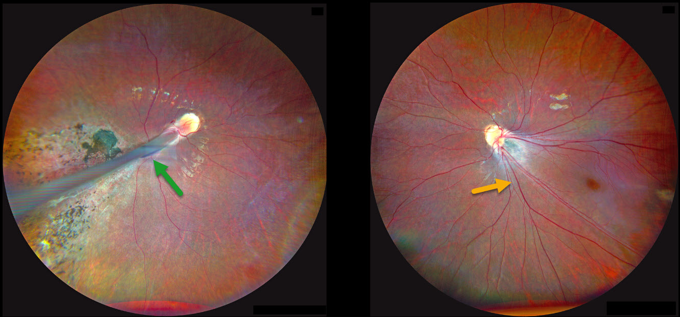
Fundus photography OD shows a prominent retinal fold extending from the optic disc into the inferotemporal periphery. Diffuse chorioretinal scarring is noted surrounding the retinal fold. The retinal vessels OS are dragged inferotemporally with marked foveal ectopia.
Learning Points:
Retinopathy of prematurity (ROP) is a potentially blinding vasoproliferative disease occurring in premature infants. Although multifactorial, current screening guidelines are based primarily on low birth weight and gestational age (Kim et al, Surv Ophthalmology 2018;63:618-637). Screening and treatment for these eyes is extremely subspecialized, with treatment options primarily including timely scatter laser photocoagulation and intravitreal anti-VEGF injections (Yonekawa et al, Retina 2017:37:2208-2225).
Patients with ROP have an increased risk for vision-threatening complications throughout their life, including vitreous hemorrhage and retinal detachment (Hsu et al, Ophthalmology Retina 2023;7:72-80). We will therefore monitor our patient yearly.



