

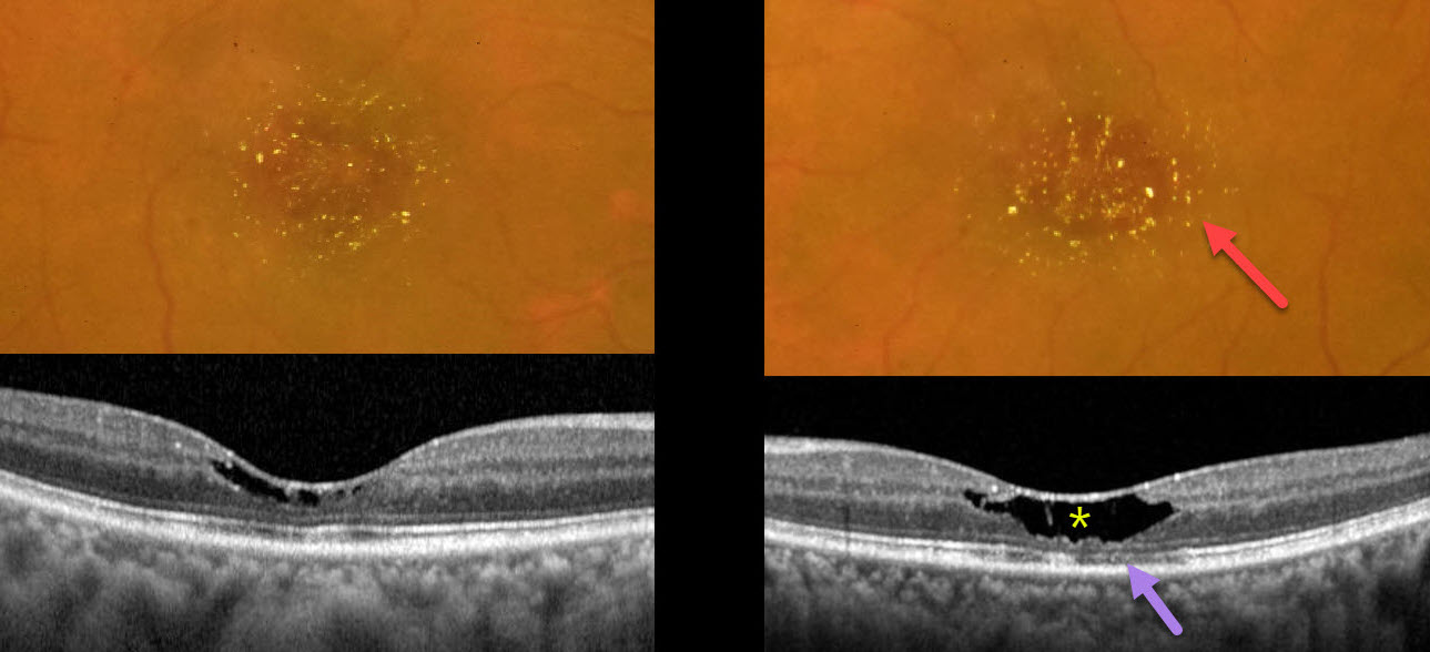
This 51YO female presented with 3 months of bilateral vision loss. She was diagnosed 5 years earlier with breast cancer, followed by a radical mastectomy. Since then, she has been treated with tamoxifen 20mg PO daily. Vision was 20/60 OD and 20/40 OS.
Color photography shows bilateral, symmetric, central macular yellowish crystalline deposits, corresponding to tiny hyperreflective inner retinal lesions on OCT. Central inner retinal cavitations, outer nuclear layer loss, and disruption of the ellipsoid zone and outer segment bands are noted, particularly in the left eye.
Learning Points:
The prevalence of tamoxifen retinopathy for patients taking tamoxifen 20mg daily may be as high as 12% (Kim et al, Ophthalmology 2020;127:555-557). Although most oncologists do not require this, it is not unreasonable to perform ophthalmic screening examinations with OCT following 2 years of therapy (Tenney et al, Surv Ophthalmology 2023;69:42-50).
Tamoxifen inhibits the glutamate-aspartate transporter, leading to excessive intracellular accumulation of glutamate in Müller cells, which are vital in maintaining retinal cell integrity and homeostasis. Tamoxifen retinopathy shows findings very similar to macular telangiectasia type 2 (MacTel 2; Lee et al., Ophthalmology Retina 2020;3:681-689), including retinal cavitations, right-angle venules, and inner retinal crystals. Both disorders likely share Muller cell injury as a common etiology. The changes in MacTel2 are usually in the temporal fovea, whereas they seem to be more diffusely distributed throughout the central macula with tamoxifen (Hess et al, Retina 2023;7:101-110).
Ocular toxicity is more common with a cumulative dose over 100 grams, although our patient still developed classic toxicity despite a cumulative dose of only 35.2 grams. The tamoxifen was discontinued after discussion with the treating oncologist. Three months later, the vision and retinal findings were unchanged (not shown).



