

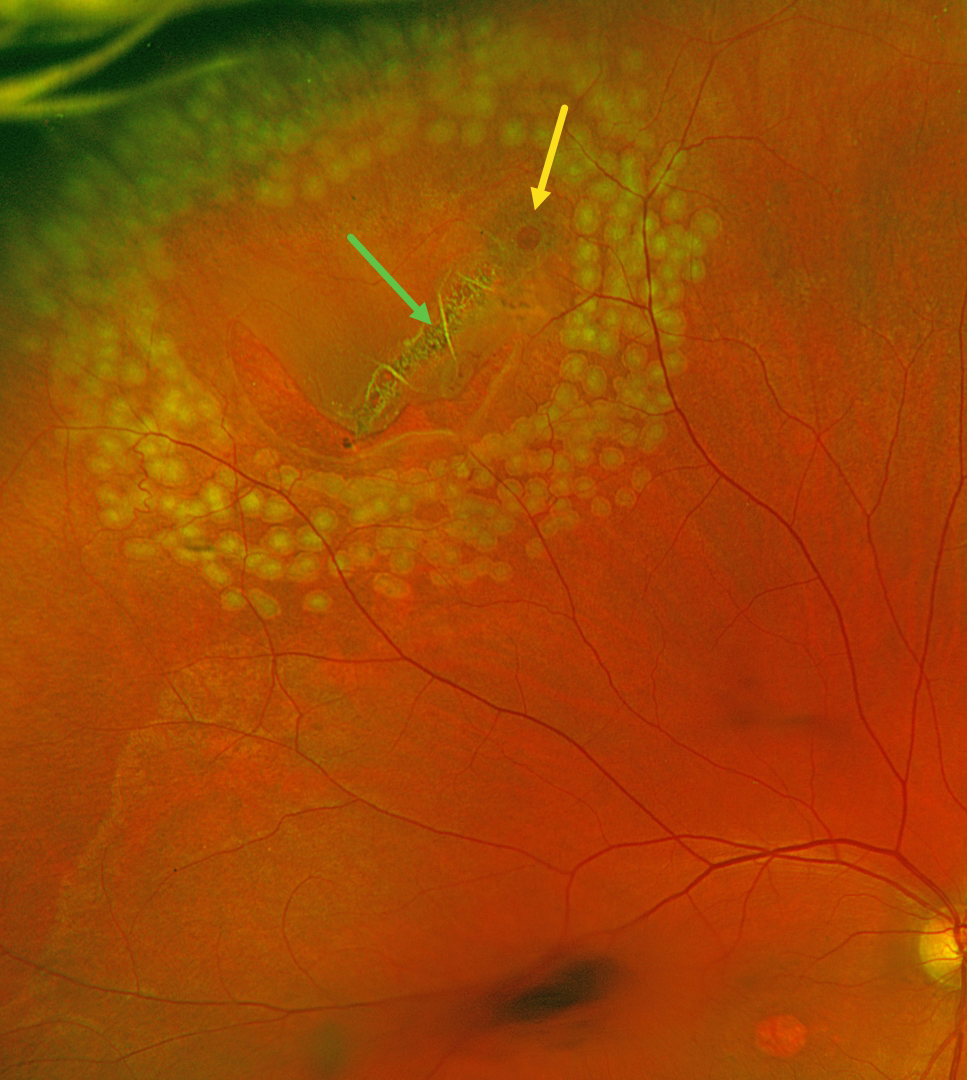
This 63YO male with history of idiopathic central serous chorioretinopathy (ICSC) with secondary macular neovascularization OD and lattice degeneration OU presented with new floaters in his right eye. He was scheduled for an anti-VEGF injection for the right eye on a treat-and-extend protocol the same day. Vision was stable at 20/100 OD and 20/20 OS. An acute, related retinal tear was found and immediately demarcated with thermal laser photocoagulation.
Optos color RG imaging immediately following the laser procedure shows the retinal break well surrounded by multiple nearly confluent rows of laser burns. The flap of the tear consists of an entire lattice lesion along with a chronic atrophic hole with surrounding pigment. There is an area of central macular atrophy from the ICSC.
Learning Points:
Lattice lesions have a pocket of liquefied vitreous overlying thinned retina with a firm area of surrounding vitreoretinal adhesion. White, fibrosed vessels often crisscross the lesions, which gives the lesions their name, “lattice.” This firm vitreoretinal adhesion, along with the often-present atrophic holes, is what can predispose these eyes to retinal breaks and detachment. Although prophylactic treatment is rarely needed, symptomatic breaks, as in our patient, need prompt retinopexy.



