

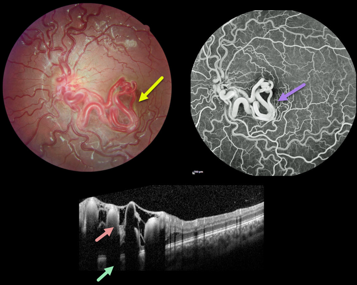
This healthy 11YO girl was seen for a routine ophthalmological examination. Vision was 20/20 OU.
Fundus photography of her left eye shows unilateral tangles of markedly dilated and tortuous vessels extending from the disc into the inferior macula and fovea. The retinal vessels elsewhere are also somewhat dilated and tortuous. OCT scanning through the inferior macula shows these hyperreflective vessels to variably extend through the full-thickness retina with posterior shadowing.
The early-phase fluorescein angiogram shows rapid filling of the dilated artery and vein, with no intervening capillaries (not shown). The late phase shows no angiographic leakage.
Learning Points:
A retinal arteriovenous malformation (AVM) is a congenital connection between an artery and vein without an intervening capillary network. These lesions appear along a spectrum ranging from small isolated lesions to the large ‘bag of worms’ appearance as seen in our patient.
These more extensive lesions are usually part of the Wyburn-Mason Syndrome, which includes vascular lesions in the eyes and midbrain. Retinal venous malformations, also known as retinal macrovessels, are now thought to represent an AVM (Pichi et al., JAMA Ophthalmol 2018;136:372-379).
Fortunately, our patient’s MRI was normal. The AVM should likely remain stable throughout life and should not require any treatment.



