

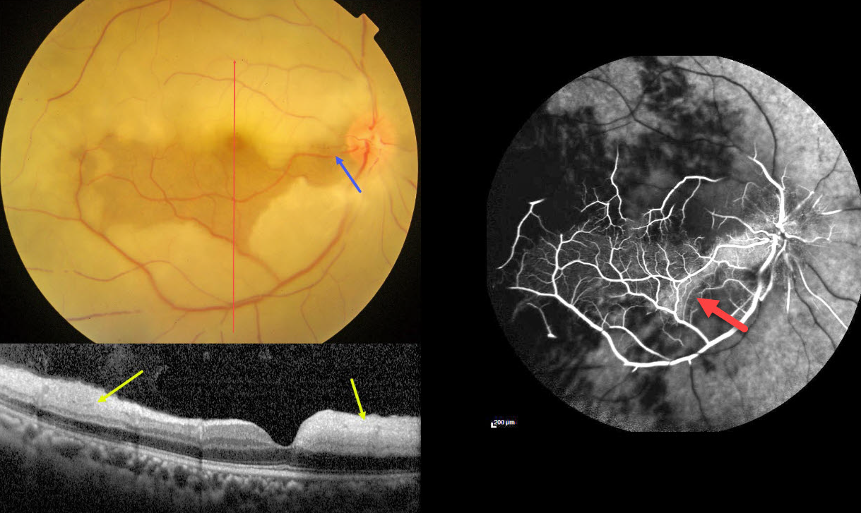
This 45YO male presented with acute vision loss in his right eye 4-5 days earlier. Vision was 20/80 in his right eye and 20/20 in his normal left eye.
Color photography shows an acute central retinal artery occlusion (CRAO) with sparing of the central and inferior macula due to a large cilioretinal artery.
The vertical OCT B-scan shows opaque hyperreflective paracentral inner retina with normal central and inferior foveal layers. Fluorescein angiography confirms preserved central and inferior macular arterial flow.
Workup elsewhere included a normal brain MRI and cardiac 2D Echo. Carotid Doppler showed a plaque in the right internal carotid artery.
Learning Points:
The inner two-thirds of the neurosensory retina is supplied by the central retinal artery, with the choroid supplying the RPE and photoreceptor layer. Acute CRAO, therefore, presents with a white edematous inner retina that spares the fovea.
Cilioretinal arteries, which arise from the posterior choroidal circulation, are present in up to 50% of individuals, and bilateral in about 25% (Schneider et al, Acta Ophthalmologica 2021;99:e310-e318). Our patient was quite fortunate to have a large cilioretinal vessel that relatively preserved central vision.
Patients with acute retinal ischemia (defined as transient monocular vision loss, acute BRAO, or acute CRAO) need to be emergently referred to a stroke center. This is especially urgent with an acute CRAO since about 75% of patients have already developed a recent stroke.
If the patient presents immediately after occlusion, measures to acutely lower intraocular pressure to move the causative embolus downstream can be performed, including ocular massage, paracentesis, and breathing into a bag. Unfortunately, most patients present outside this window (Shah et al, Ophthalmology Retina 2023;7:527-531), and even then, these measures are usually ineffective.



