

This 41YO female presented with headaches, redness, and pain OD for 1.5 months and OS for 2 weeks. She was diagnosed elsewhere with angle closure glaucoma, received a YAG peripheral iridotomy, and started on glaucoma drops. She then saw multiple other doctors who diagnosed her with papilledema.
Her CSF opening pressure was 21, and she was started on oral acetazolamide for a presumed diagnosis of idiopathic intracranial hypertension (IIH). In our office, vision was 20/100 OD and 20/60 OS. Intraocular pressure (IOP) was 30mmHG.
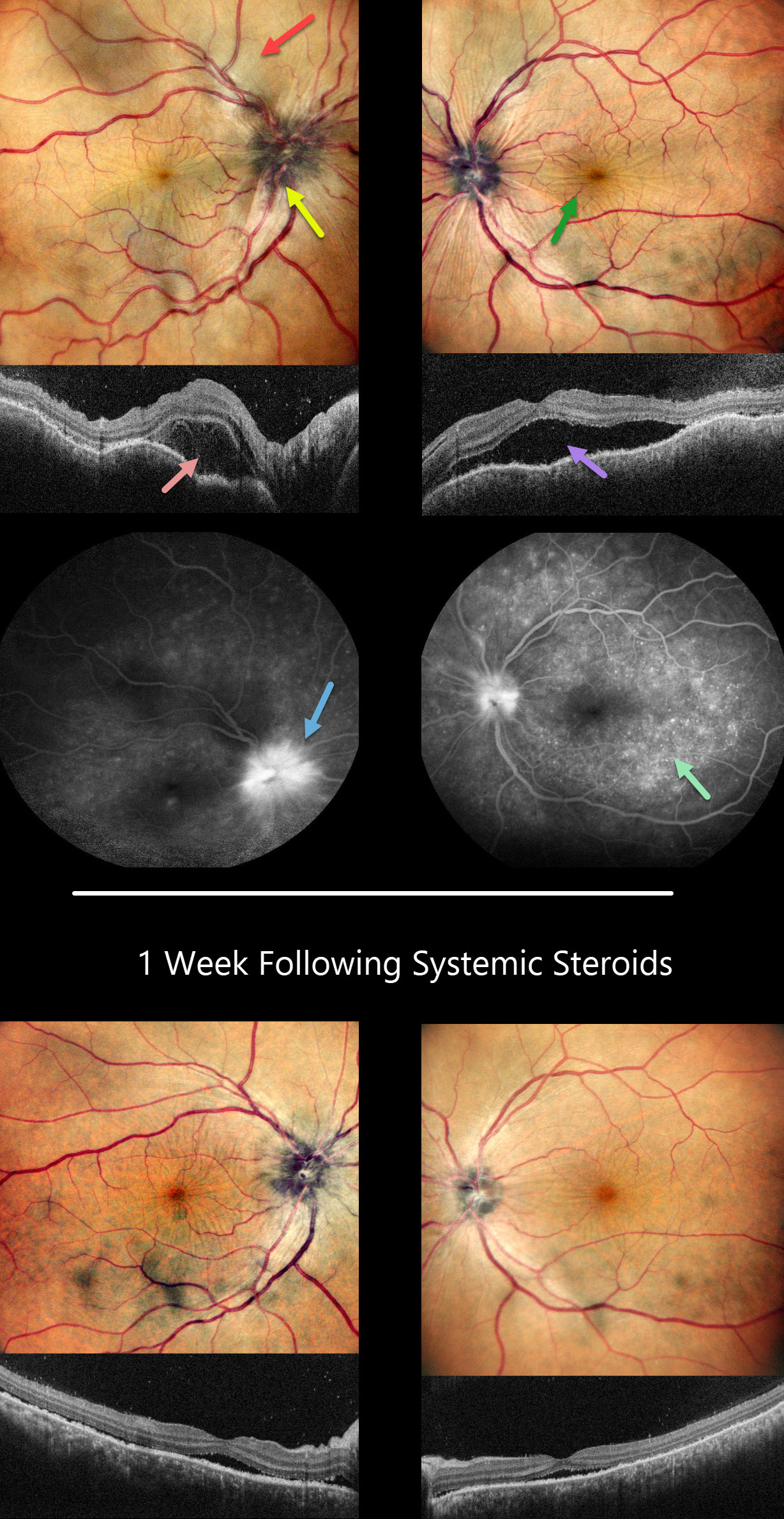
Anterior segments showed shallow anterior chambers with retrolental vitreous cells. Multicolor imaging shows bilateral irregular chorioretinal folds, multifocal serous detachments, and disc hyperemia.
OCT scanning shows a bacillary layer detachment OD, subretinal fluid OS, and a bilateral undulating thickened choroid. Fluorescein angiography shows bilateral optic nerve involvement and pinpoint subretinal leakage.
She was diagnosed with Harada’s disease and started on intravenous methylprednisolone for 3 days, followed by 60mg prednisone PO daily. She was also referred to rheumatology to start immunosuppressants. The acetazolamide was tapered and discontinued for the misdiagnosed IIH.
One week later, vision improved to 20/40 OU. IOP was 10mmHG OU with marked bilateral improvement in the chorioretinal folds, macular fluid, and choroidal thickening. A slow steroid taper was initiated pending initiation of immunosuppressants.
Learning Points:
This case has many classic findings for Harada’s disease, including bilateral panuveitis, optic nerve swelling, thickened choroid, chorioretinal folds, and multifocal serous exudative retinal detachments.
Patients with just ocular findings have Harada’s disease, whereas those with additional systemic findings (including vitiligo, poliosis, headache, vertigo, and hearing loss) have Vogt-Koyanagi-Harada (VKH) disease.
This case also reminds us that it is easy to misdiagnose these complex uveitis cases, especially when not all ocular findings are taken into full consideration in an attempt to give the patient a single, unifying diagnosis.



