

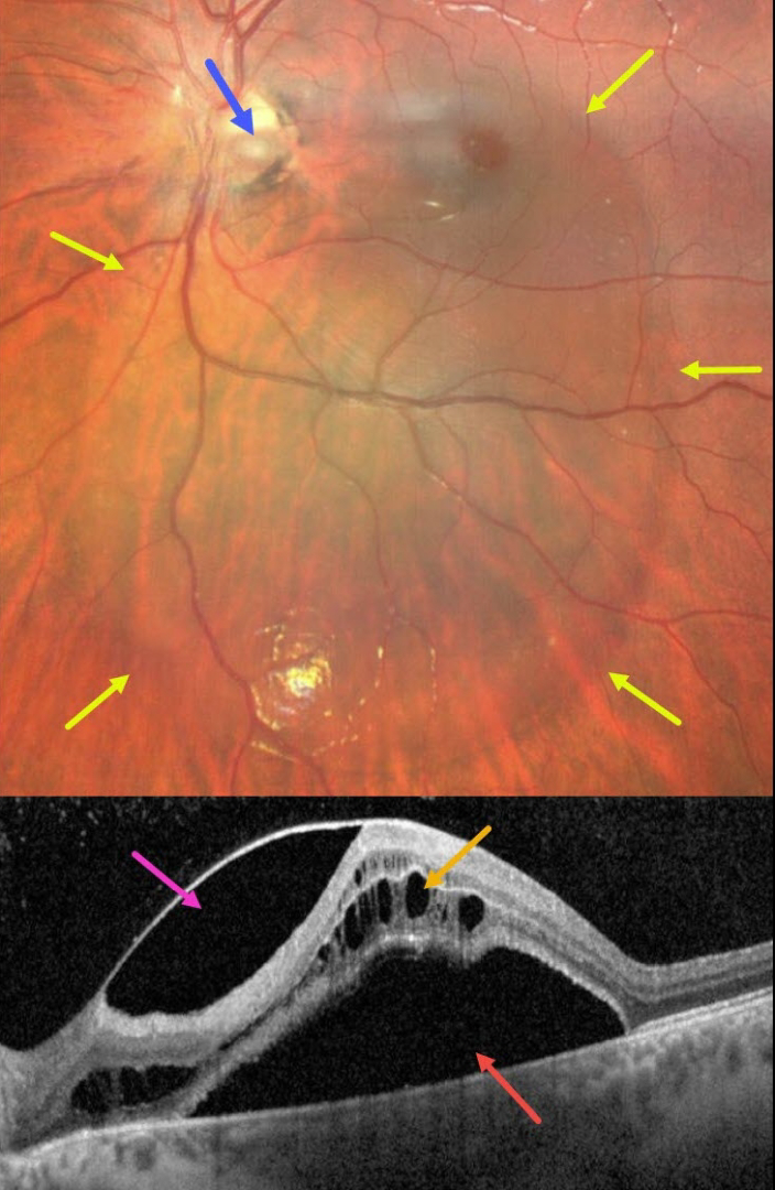
This 14YO boy presented with these asymptomatic and unilateral findings in his left eye. Vision was 20/50.
Fundus imaging shows a large vertical oval of retinal elevation extending from the superior macula into the inferior midperiphery. This fluid connects to an inferotemporal optic nerve pit.
OCT scanning shows subretinal fluid, cystic fluid mostly in the outer nuclear layer, and fluid below the internal limiting membrane vs within a split nerve fiber layer. He was referred to a retinal specialist for further evaluation and possible treatment.
Learning Points:
An optic nerve pit is a rare (about 0.1% prevalence) congenital anomaly thought to be caused by incomplete closure of the optic fissure during gestation. Pits are most commonly located within or along the inferior-temporal disc margin and usually appear as a gray depression.
While most optic pits are asymptomatic (as in this case), they can cause vision loss by forcing fluid into the inner retina, outer retina, and, ultimately, the subretinal space (optic pit maculopathy). The source of the fluid remains a subject of ongoing debate, with opinions ranging from the vitreous to the subarachnoid space.
A host of treatments for optic pit maculopathy have been suggested over the years. However, a recent meta-analysis suggested that a plain-vanilla vitrectomy without gas tamponade is as successful as other procedures (Zheng et al., Ophthalmology Retina 2020;4:289-299).



