

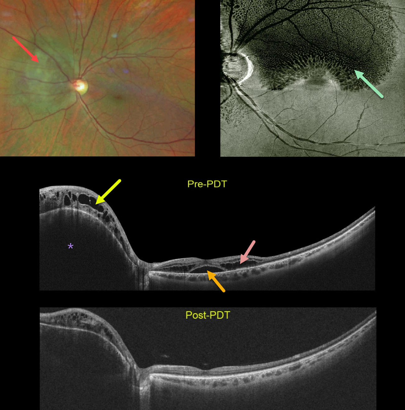
This healthy 45YO female was referred for macular schisis. Vision was 20/50. Color photography shows an orange-red subretinal lesion nasal to the nerve.
Widefield OCT shows an elevated hyporeflective choroidal mass with overlying cystic retinal edema. Subfoveal fluid with cystic fluid in the outer retina is noted in the macula. An en face OCT of the outer retina more dramatically shows the cystic changes.
The choroidal lesion with overlying retinal edema dramatically improved following photodynamic therapy. Vision improved to 20/20.
Learning Points:
Widefield imaging is revolutionizing the way we diagnose and treat retinal disease. This case is a perfect example in which macular imaging alone could lead to an incorrect diagnosis.
In this case, the macular findings in isolation suggest either optic pit maculopathy (Imamura et al, Retina 2010;30:1104-1112) or paraproteinemic maculopathy (Mansour et al, Ophthalmology 2014;121:1925-1932). This widefield technology led to a correct diagnosis, allowing for appropriate treatment with excellent anatomic and visual results.



