

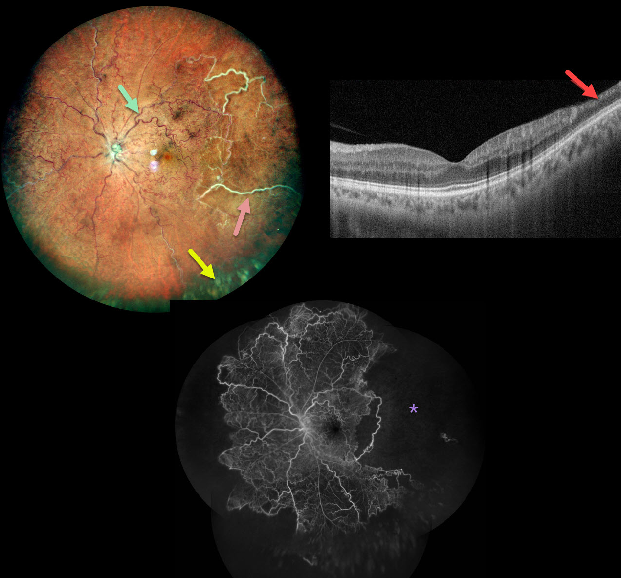
This healthy 34YO male presented with this relatively asymptomatic central retinal vein occlusion (CRVO). Vision was 20/20 OU.
Color imaging shows large temporal nonperfused retinal vessels with old inferior yellow vitreous hemorrhage. The retinal vessels are diffusely dilated and tortuous. OCT B-scan shows a remarkably normal central macula with some distal inner retinal thinning. Fluorescein angiography confirms widespread peripheral nonperfusion.
Anti-VEGF therapy, followed by panretinal photocoagulation, was administered.
Learning Points:
The Central Vein Occlusion Study (CVOS) defined ischemic CRVO as having at least 10 disc areas of angiographic retinal nonperfusion as determined by standard photographic views with the Canon (60 degrees) or Topcon (45 degrees) wide-angle fundus cameras (Ophthalmology 1995;102:1434-1444).
Although clearly ischemic, this patient might have been classified by the CVOS as a perfused occlusion, depending on the extent of peripheral ischemia imaged. As with diabetic retinopathy (see Brucker, Retina 2021;41:461-463), ultrawidefield imaging and anti-VEGF injections have thrown a literal wrench into how we think about, classify, and treat retinal disorders.



