

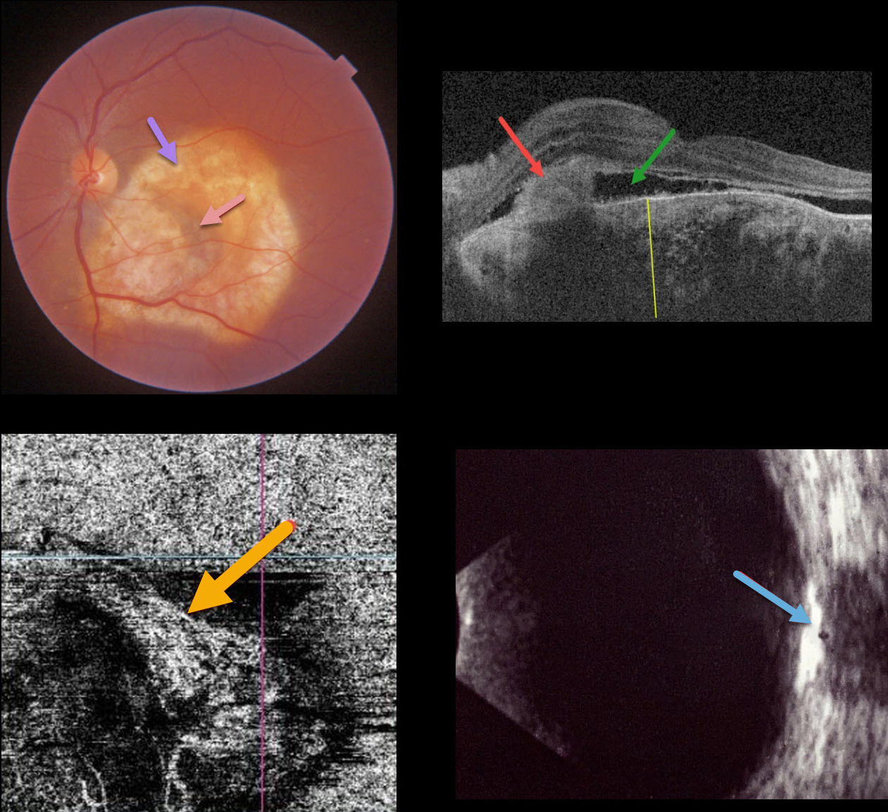
This 40YO female presented with recent vision loss in her left eye. Vision was 20/20 in her normal right eye and counting fingers in her left eye.
Triton color imaging shows an amelanotic choroidal lesion occupying virtually the entire macula. There is a small dot of central blood. Swept-source OCT shows a markedly thickened choroid with variable regions of hyper- and hyporeflectivity. A hyperreflective type 2 macular neovascularization (MNV) is seen along with exudative subretinal fluid. OCT angiography shows flow signals within the MNV. B-scan ultrasonography shows the lesion to be markedly hyperreflective.
An anti-VEGF injection was given, but she was unfortunately subsequently lost to follow-up.
Learning Points:
Choroidal osteomas are benign, calcified amelanotic choroidal tumors. They are more common in women, are almost always unilateral, and are usually located in the peripapillary or macular regions.
Choroidal lesions are best seen on the Optos red channel. Fluorescein angiography (FA) usually shows early patchy hyperfluorescence with late diffuse staining. Ultrasonography is often helpful in differentiating an osteoma (markedly hyperreflective) from other amelanotic choroidal tumors.
Macular neovascularization develops in a significant number of eyes and can be successfully treated with anti-VEGF therapy (see Seong et al, Graefe’s 2022;250:1713-1721).



