

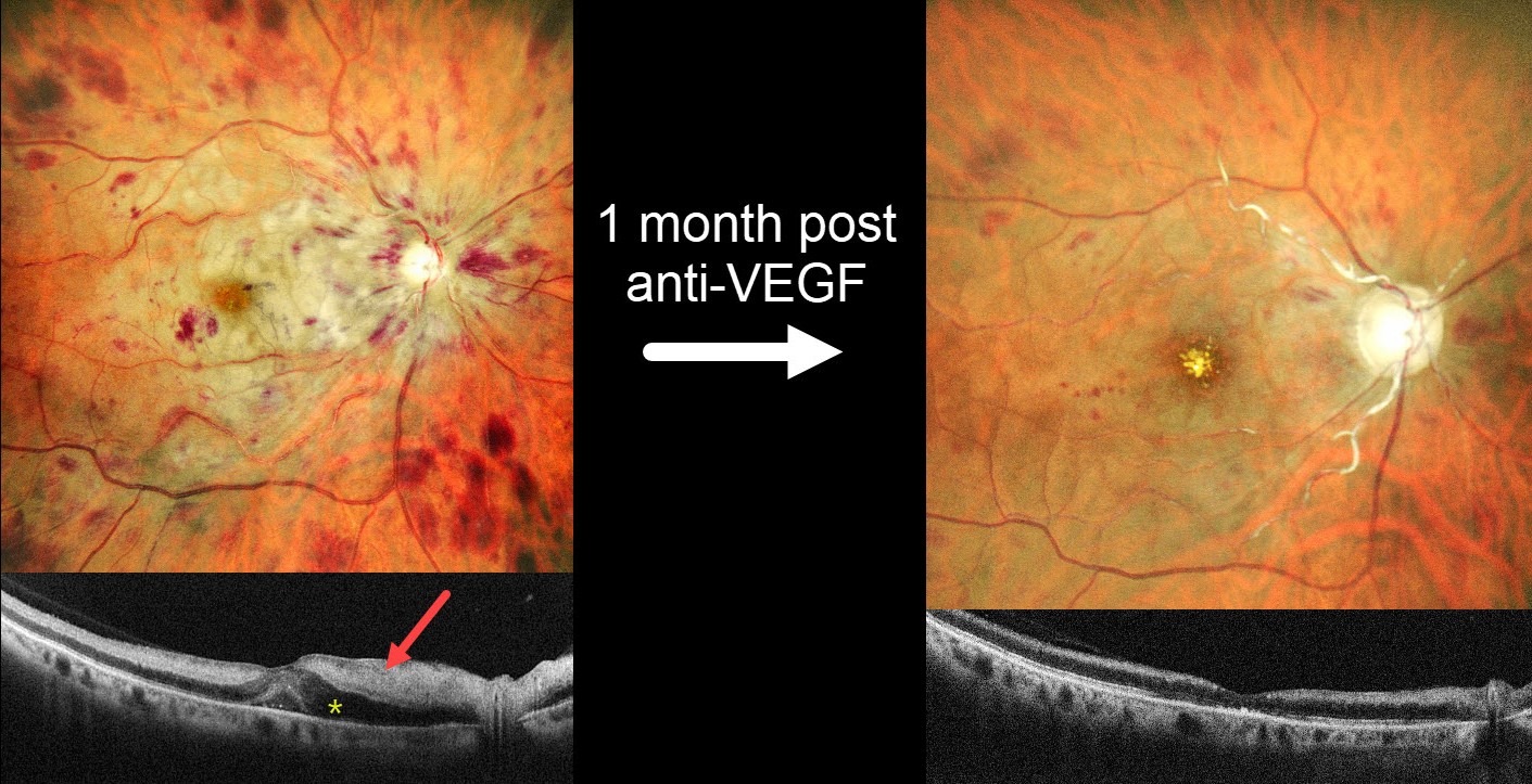
This 57YO female with a history of severe hypertension presented with one day of severe vision loss in her right eye. Blood pressure was 200/100 mmHg. Vision was hand motion OD and 20/20 in her normal OS.
MultiColor SLO imaging shows inner retinal opacification sparing the macular center, consistent with a fresh central retinal artery occlusion (CRAO). A central retinal vein occlusion (CRVO) is also noted with a swollen nerve and retinal hemorrhages in all quadrants. OCT scanning shows marked hyperreflectivity with loss of the band detail throughout the inner and mid-retinal layers. The peripapillary retina is edematous with fluid in the nasal outer nuclear layer and mild foveal subretinal fluid.
She was diagnosed with a combined CRAO-CRVO. An intravitreal anti-VEGF injection, and 1 month later, vision improved to 20/400. Repeat imaging shows marked improvement in the retinal hemorrhages, retinal opacification, and macular fluid, although new foveal lipid is noted. The nerve is flat and pale, and the major proximal arterioles are white without a blood column. The inner retina remains hyperreflective on OCT but is thinning with loss of the normal architecture. Panretinal photocoagulation was scheduled.
Learning Points:
Combined CRAO-CRVO is thankfully an extremely rare event, accounting for 0.3% of all retinal vascular occlusions (Raval et al, Indian J Ophthalmology 2020;68:2136-2142). Risk factors appear similar to those associated with isolated occlusions. The CRAO may be embolic or secondary to compression from a swollen optic nerve.



