

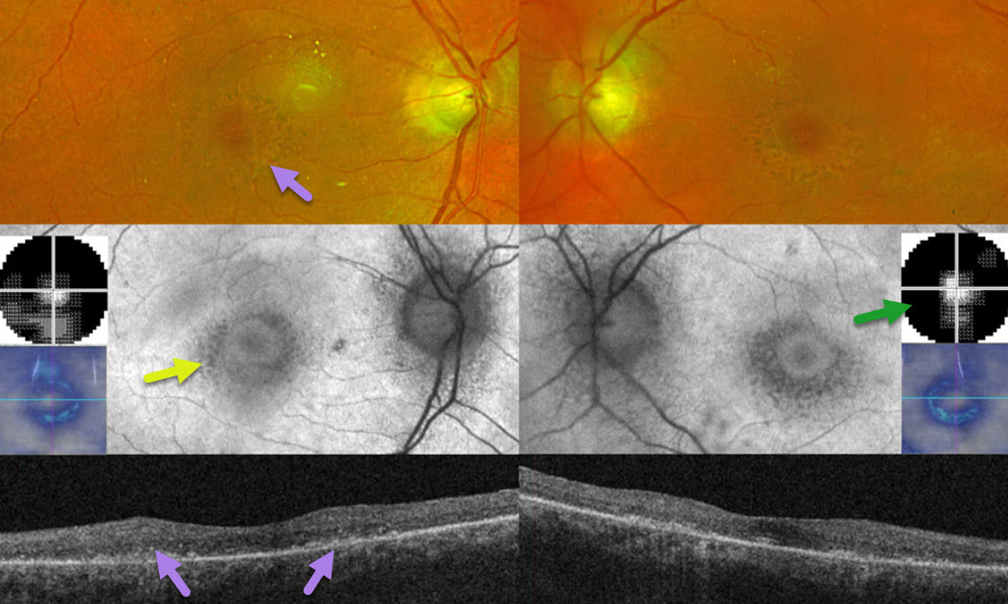
This 78YO female was on Plaquenil (hydroxychloroquine) for 7 years for her “arthritis.” After noticing progressive reading difficulties, she decided to stop this medicine about 3 months earlier. She was screened annually by her local eye doctor, who referred her for possible toxicity. Vision was 20/40 OD and 20/30 OS.
Optos color imaging and OCT show classic findings for severe hydroxychloroquine toxicity, including an oval area of foveal pigment loss/mottling and outer retinal atrophy. These areas of pigment loss hypo-autofluoresce. 10-2 visual fields reveal small islands of preserved central vision.
Learning Points:
Current screening guidelines should make Plaquenil toxicity a thing of the past. The American Academy of Ophthalmology updated its screening guidelines in 2025 (Marmor et al, Ophthalmology 2026;133:439-450).
A baseline screening examination with fundus photography, spectral-domain or swept-source OCT, and fundus autofluorescence (FAF) should be performed as soon as treatment is started to note preexisting conditions and for future comparison. Annual screening is recommended but may be deferred during the first 5 years if there are no significant risk factors, ie, high daily dosage, renal disease, concomitant drugs such as tamoxifen, macular disease, and older age.
The recommended daily dosage remains unchanged at <=5.0 mg/kg/day real weight, with the dosage under 400 mg/day for severely obese individuals. The primary screening tools are OCT with widefield FAF. Secondary confirmatory tests include 24-2C visual field testing (which screens the parafoveal and pericentral regions simultaneously) and multifocal ERG. The pericentral field needs to be evaluated, as Asians typically have more eccentric macular and midperipheral involvement.



