

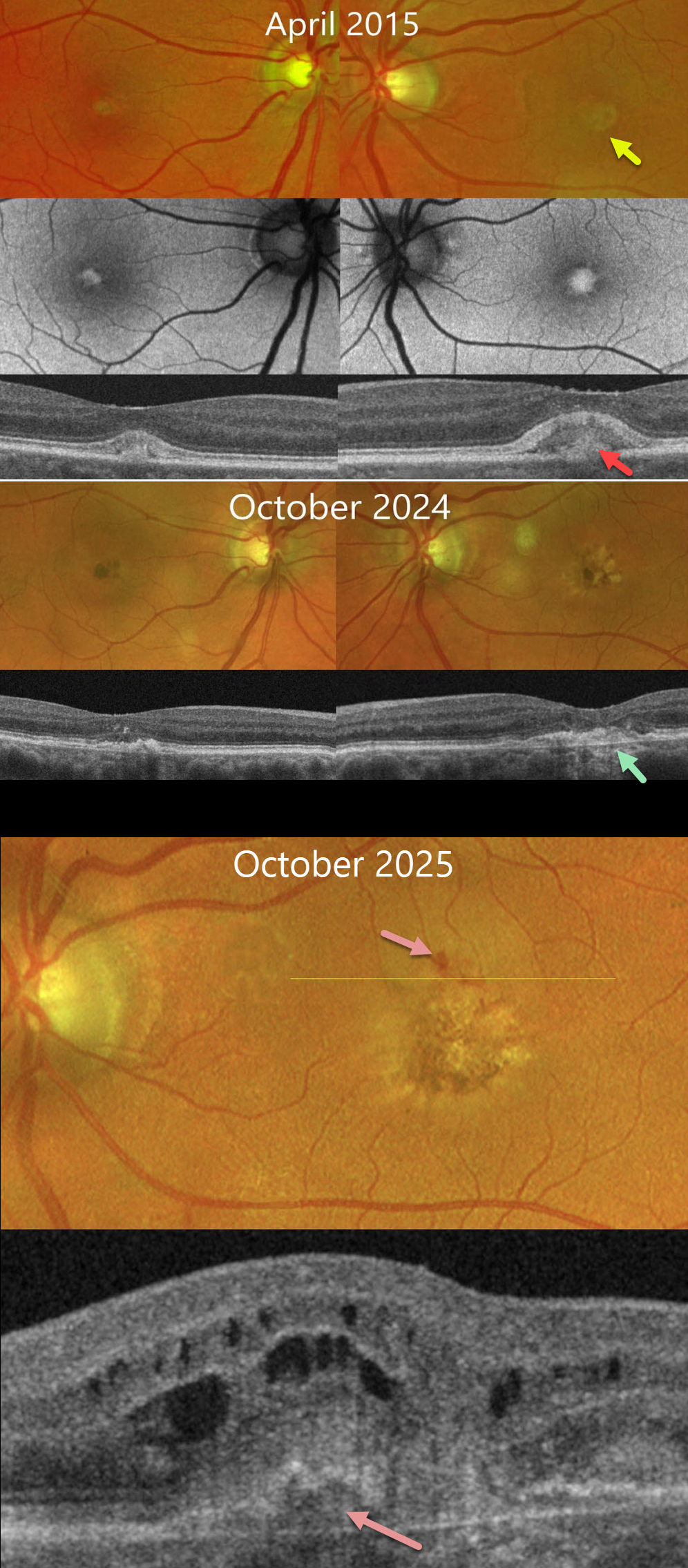
This 74YO female was referred to us in April 2015 for asymptomatic age-related macular degeneration. Vision was 20/20 OD and 20/25 OS.
Fundus photography shows bilateral foveal yellowish pigmented lesions consistent with adult-onset foveomacular vitelliform dystrophy, which hyper-autofluoresce. On OCT, these appear as focal subfoveal hyperreflective lesions that indent the overlying retina. She was referred back to us in October 2024 with a gradual symptomatic decrease in vision to 20/40 OD and 20/70 OS. There is increased foveal pigmentation with collapse of the vitelliform lesions on OCT.
She returned in October 2025 with an acute change of vision in her left eye. Vision was 20/200, with a new macular neovascularization (MNV) and macular fluid and blood noted on funduscopic exam and on OCT. Following 2 monthly intravitreal Vabysmo injections, vision improved to 20/100 with resolution of OCT fluid (not shown).
Learning Points:
Adult onset foveomacular vitelliform dystrophy is characterized by bilateral, symmetrical, grayish-yellow, one-third to one-half disc diameter subfoveal lesions with a central pigmented spot. In our experience, it is the most common pattern dystrophy and resembles Best disease, although the lesions are generally smaller and diagnosed in adulthood. Causative mutations include BEST1, PRPH2, IMPG1 and IMPG2. For a great review article, see Chowers et al, Progress in Retinal and Eye Research 2015;47:64-85.
MNV in adult-onset foveomacular dystrophy has been reported to occur in 5-15% of eyes (Da Pozzo et al, Ophthalmologica 2001;215:412-414). Patients, therefore, need to be examined at least yearly. MNV usually responds well to anti-VEGF therapy (Mimoun et al, Retina 2013;33:513-521).



