

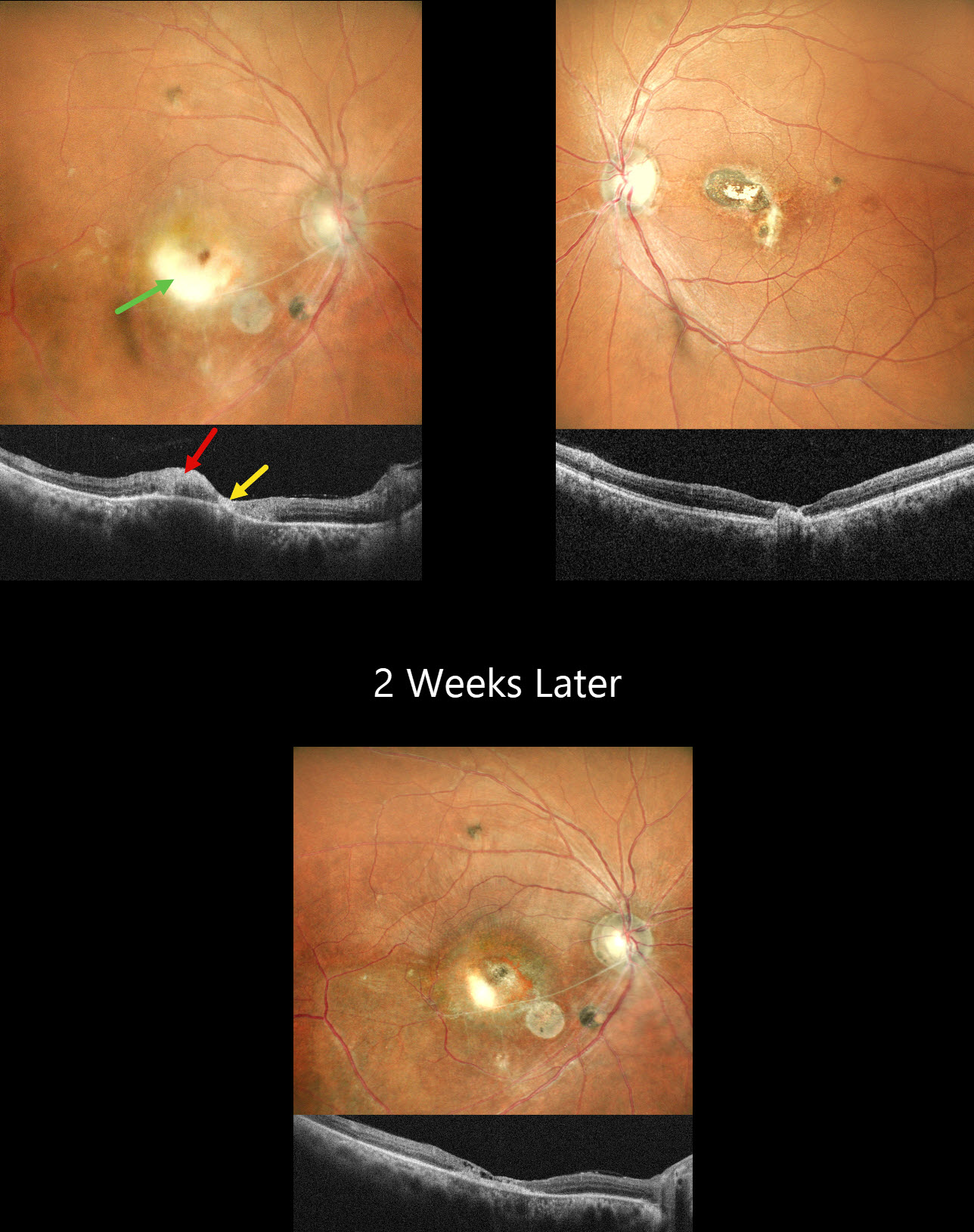
This healthy 45YO male presented with a 3-week history of decreased vision in his right eye. He reported a similar episode in the left eye 10 years earlier, with poor vision since. Vision was 20/120 OD and 20/200 OS.
Pseudocolor SLO imaging OD shows active white retinitis adjacent to inactive nasal scarring. The vessels appear sclerosed in the inferonasal macula. OCT shows hyperreflectivity within the area of active retinitis with atrophy within the nasal scarring. The left eye has inactive nasal macular scarring. He was diagnosed with recurrent toxoplasmosis and started on a 6-week course of oral Bactrim DS. Oral steroids were also started 3 days later. When he returned 2 weeks later, he was unchanged at 20/120 with resolution of the active retinitis.
Learning Points:
Toxoplasmosis, the most common cause of infectious chorioretinitis in humans, is caused by the protozoan parasite, Toxoplasma gondii. It is usually transmitted through infected felines or by eating raw meat. Most infections are acquired, although it can also be transmitted congenitally. The body’s immune system is unable to completely kill the organism, which often lies dormant within a chorioretinal scar for years. When the immune balance favors the encysted organism, active chorioretinitis develops. Focal retinitis usually develops adjacent to a pigmented retinochoroidal scar. Overlying retinal arterial vasculitis is not uncommon.
The inflammation usually resolves spontaneously within 6 weeks. If the optic nerve or macula are threatened, as in our patient, a six-week course of Bactrim DS is recommended (Soheilian et al, Ophthalmology 2004;112:1876-1882). Oral prednisone can be added as well to help with severe inflammation.



