

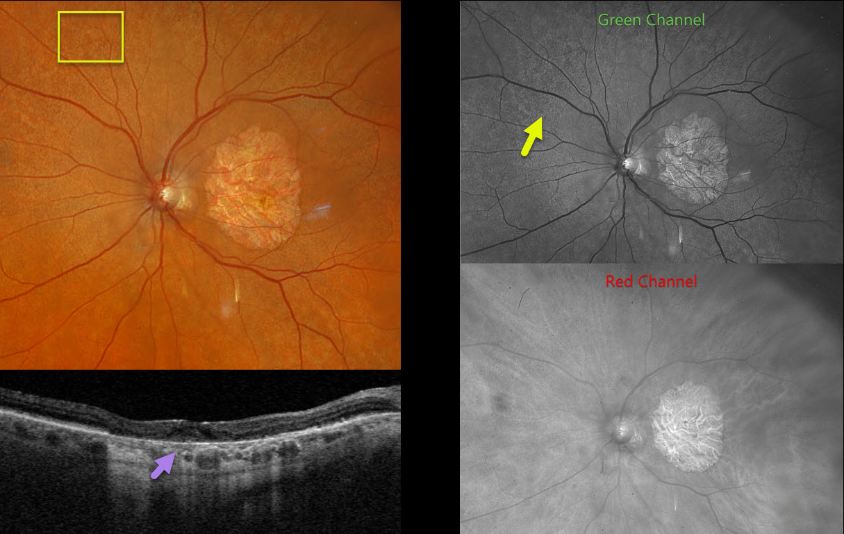
This 78YO female, previously diagnosed with macular atrophy from age-related macular degeneration, has stable vision of 20/400 OD and counting fingers OS.
Optos color RGB imaging of her left eye shows extensive subretinal drusenoid deposits scattered throughout the posterior pole. These are best seen in the green channel. Triton swept-source OCT shows variable incomplete outer retinal and RPE atrophy. Identical findings were noted in her right eye (not shown).
Learning Points:
Originally described by Hamel et al (AJO 2009;147:609-620), extensive macular atrophy with pseudodrusen (EMAP) is an idiopathic, rapidly progressive macular atrophy. Unlike age-related macular atrophy, EMAP begins at an earlier age (50-55 years) and progresses more rapidly to legal blindness within 5 years. The macular atrophy is typically greater in the vertical meridian. Patients also have extensive subretinal drusenoid deposits (reticular pseudodrusen) with extensive peripheral cobblestone degeneration.
For an excellent recent review, see Antropoli et al, Progress in Retinal and Eye Research 2025;104:101320.



