

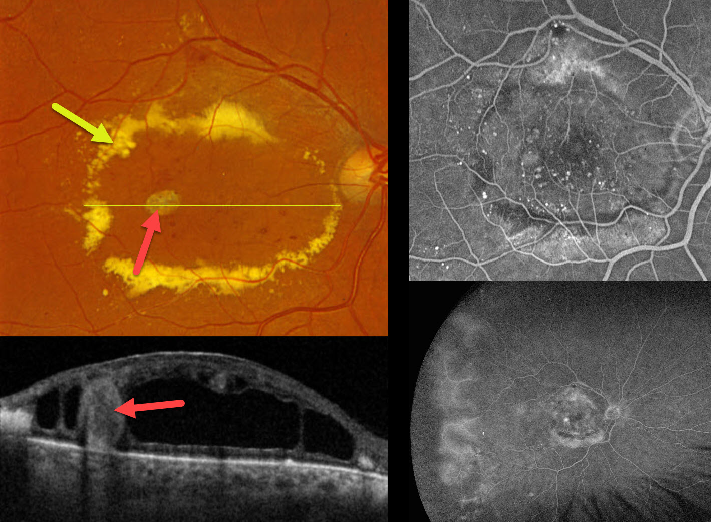
This 71YO male presented with a vague history of decreased vision in his right eye. He gave a history of diet-controlled type 2 diabetes. Vision was 20/200 OD and 20/20 in his normal OS.
Optos color RGB imaging shows an oval ring of subretinal lipid surrounding the central macula. Scattered telangiectatic vessels are noted. A large fibrosed macroaneurysm is seen temporally. On Triton swept-source OCT, this macroaneurysm spans nearly the entire retinal thickness and is surrounded by a large mid-retinal cystic cavity. Fundus fluorescein angiography (FFA) shows numerous macular microaneurysms with relative blockage from the subretinal lipid. There is diffuse leakage from the temporal vessels with ischemic retina more peripherally associated with some telangiectatic vessels. FFA was normal in his left eye (not shown). Intravitreal anti-VEGF therapy was started.
Learning Points:
Described by George Coats in 1908, Coats disease is an idiopathic disorder, usually unilateral, in males. It is characterized by retinal vascular telangiectasia involving capillaries, arterioles, and venules. Patients can present with a wide range of findings, from minimal exudation to total exudative retinal detachment. The leaking vessels are usually treated to prevent exudative complications.
Thermal laser photocoagulation is usually the initial treatment modality, but anti-VEGF injections can also be used to stabilize the vascular leakage. Treatment involves a combination of focal laser to the leaking vascular abnormalities along with scatter laser into areas of ischemia. Patients will often require multiple rounds of treatment. See Sigler et al for a review of the current management (Survey Ophthalmology 2014;59:30-46).



