

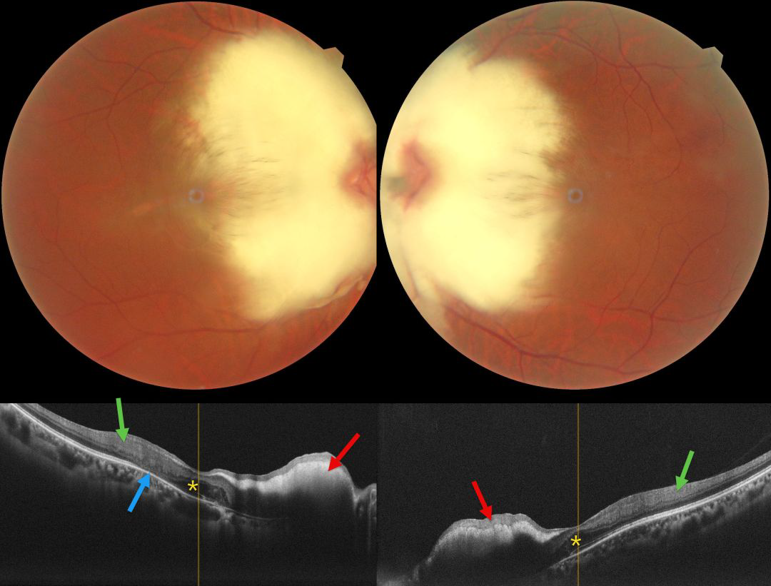
This healthy 36YO female presented with high myopia of -11.50 -1.75 x 50 OD and -11.00 -1.00 x 170 OS. Vision was 20/80 OD and 20/60 OS. Color photography shows a bilaterally symmetric myelinated nerve fiber layer (NFL) covering each nerve and the nasal macula. The discs may also be hypoplastic, although the margins are difficult to discern due to the myelination.
On OCT, the myelination appears as a markedly hyperreflective, thickened inner retina with posterior shadowing. The foveal outer nuclear layers (ONL) appear thickened OU. There is hyperreflective material in the right nasal foveal ONL with disorganization of the underlying outer retinal bands. The inner retinal bands are disorganized more temporally OU, with outer retinal atrophy OD.
Learning Points:
Myelinated NFL occurs when retinal nerve fibers develop a myelin sheath, which usually stops posterior to the lamina cribrosa. The myelination may represent an oligodendrocytic choristoma (Rao et al, Retina 2019;39:1125-1132).
Myelinated NFL is a relatively uncommon, unilateral or bilateral, usually isolated finding present at birth. The myelinated NFL is often connected to the optic nerve. Most patients are asymptomatic, although some may have a corresponding relative scotoma. The myelination can progress in about 10% of cases and can also resolve following retinal ischemic events. Our patient’s findings were consistent with Straatsma syndrome, which consists of the triad of bilateral myopia, amblyopia, and myelinated NFL.
Myelinated NFL is reported to anatomically affect just the NFL (Rao et al, Retina 2019;39:1125-1132). Our patient’s OCT findings showing both inner and outer retinal changes outside the area of myelination has not been previously reported.



