

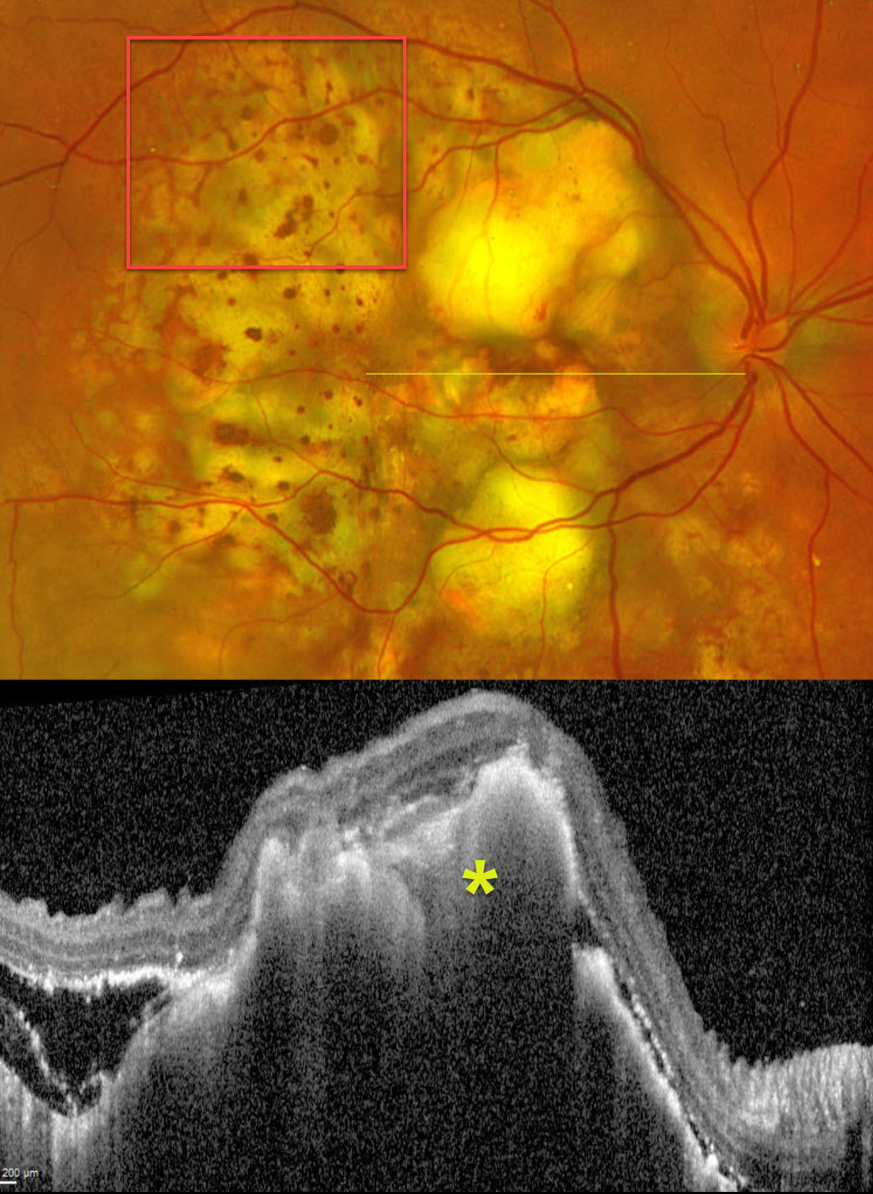
This 67YO male presented with progressive decreased vision in his right eye. He had a known history of metastatic lung cancer. Vision was hand motion OD.
Optos color RG imaging shows multifocal, variably confluent, creamy-yellow subretinal lesions scattered within the macula and extending into the midperiphery. A leopard spot pattern of pigmentary changes is noted temporally. OCT scanning shows marked hyperreflective lumpy choroidal thickening with posterior shadowing. There is temporal subretinal fluid containing variable hyperreflective material.
Learning Points:
Although uveal melanoma is the most common primary intraocular malignancy, metastatic choroidal tumors are the most common intraocular malignancies. Lung and breast cancer are the most common sources.



