

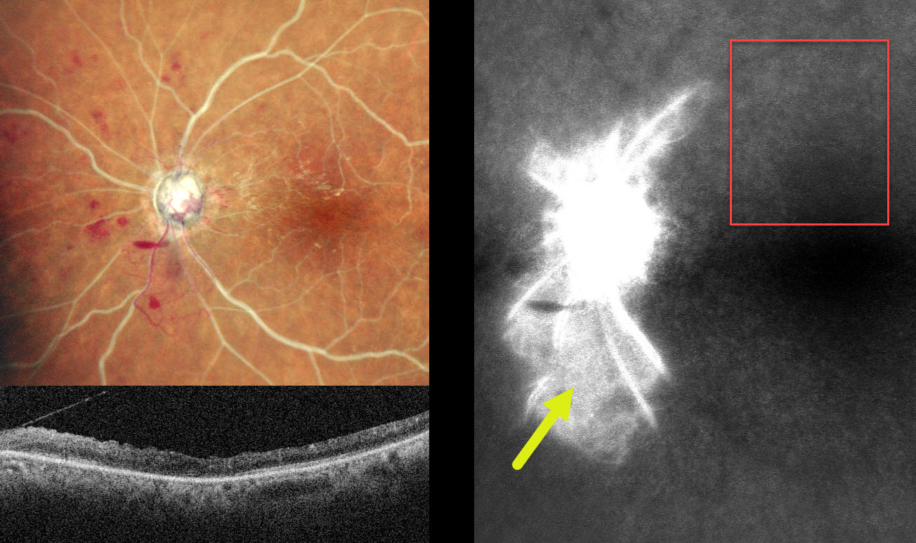
This 60YO male presented with sudden vision loss in his left eye 1 month earlier. Vision was 20/20 in his normal OD and no light perception OS.
MultiColor SLO imaging shows a combined central retinal artery occlusion (CRAO) and central retinal vein occlusion (CRVO) with white arteries and veins with no blood column. Disc neovascularization is present inferotemporally, and a small preretinal hemorrhage is noted just inferonasal to the nerve. OCT shows a disorganized hyperreflective inner retina. Fluorescein angiography shows profound panretinal ischemia with a small island of preserved capillary bed inferior to the nerve.
Vasculitis workup was negative. An emergent stroke workup, including carotid Doppler, MRI, and neurologic consultation, was ordered. Given our patient’s vision, comfort measures for his left eye were recommended.
Learning Points:
Combined CRAO-CRVO is thankfully an extremely rare event, accounting for 0.3% of all retinal vascular occlusions (Raval et al, Indian J Ophthalmology 2020;68:2136-2142). Risk factors appear similar to those associated with isolated occlusions. The CRAO may be either embolic or secondary to compression from a swollen optic nerve.



