

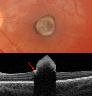
This healthy 16YO male was referred for an asymptomatic lesion in his left eye. Vision was 20/20 in his normal OD and 20/50 OS.
Triton color imaging shows a foveal hyperpigmented lesion with a nasal and superior rim of more intense pigmentation. Triton swept-source OCT shows a markedly hyperreflective intraretinal lesion that protrudes above the retinal surface with marked posterior shadowing. The funduscopic rim of increased pigment is more hyperreflective than the rest of the larger lesion. Observation was recommended.
Learning Points:
RPE hamartoma is an extremely rare, benign, presumed congenital lesion. It appears as a small, jet-black inner vs full-thickness foveal or juxta-foveal retinal lesion (Shields et al, Ophthalmology 2003;110:1005-1011). These lesions are markedly hyperreflective on OCT (Takahashi et al, Retinal Cases & Brief Reports 2013;7:179-182).



