

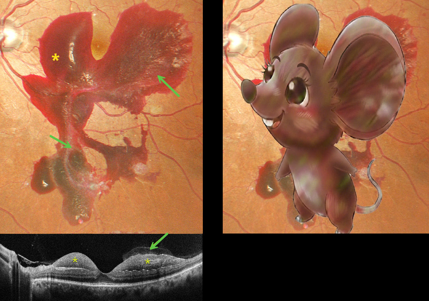
This 48YO male with a 15-year history of poorly controlled type 2 diabetes presented with one day of blurred vision in his left eye. Vision was 20/20 bilaterally.
Pseudocolor SLO imaging shows a large macular hemorrhage, sparing the foveal center, which extends into the midperiphery. The hemorrhage is mostly subhyaloid with some breakthrough blood into the vitreous. The blood compartments are well visualized on OCT.
Learning Points:
The normal vitreous undergoes a decades-long process of orchestrated degeneration, including liquefaction and gradual separation from the retinal surface. The posterior vitreous detachment (PVD) begins in the perifoveal retina early in life, followed by a foveal PVD, then a peripheral PVD, and then finally a complete PVD when the vitreous separates from the optic nerve (Johnson AJO 2010;149:371-382). Although a diabetic subhyaloid hemorrhage can extend throughout the macula, our patient’s findings illustrate the normal increase in vitreofoveal adhesion.



