

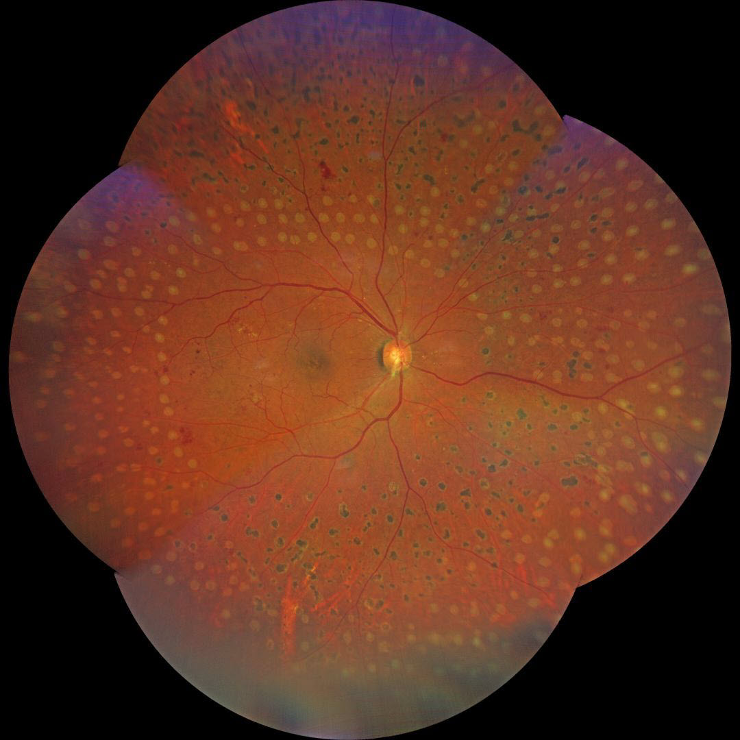
This 63YO male with type 2 diabetes presented to us with a history of recent panretinal photocoagulation (PRP) OD elsewhere. Vision was 20/20 OU.
Color photography shows irregularly sized and variably confluent laser scars mostly in the right inferior, nasal, and inferior midperiphery. Fill-in laser was applied at the slit lamp to complete the PRP.
Learning Points:
PRP should begin at least 1 disc diameter from the optic nerve and outside the major temporal arcades. Treatment is performed with a wide-angle lens or an indirect ophthalmoscope and should be applied from the posterior retina towards the periphery to prevent accidental macular burns. The laser spots should be 200-500 microns in size at the retina, be spaced about one burn-width apart, and should extend as far anteriorly as possible.
We generally do not recommend titrating the PRP based on involution of neovascularization, primarily due to the significant risk of the patient being lost to follow-up (Obeid et al, Ophthalmology 2018;125:1386-1392). Once the decision is made for PRP, we complete treatment in several sessions within a few months. Postoperative ultra-widefield imaging is helpful for assessing the adequacy of laser coverage, as areas of retinal ischemia are often undertreated (Sowmya L et al., Retina 2023;43:1922-1927).
We aren’t certain whether the initially treating laser surgeon planned further laser treatment, but the treatment was not performed in a controlled enough manner to control for burn size and spacing.



