

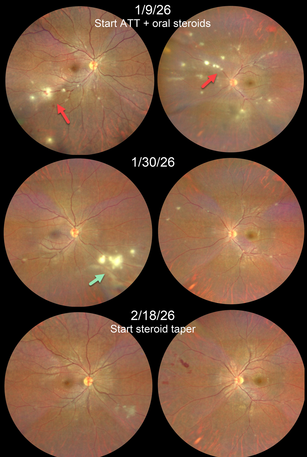
This 22YO male presented with a 1-week history of blurred vision in his left eye. His sister had been undergoing treatment for pulmonary tuberculosis 1 year ago. She also had a history of Takayasu arteritis requiring renal artery embolization and was on multiple immunosuppressants. Vision was 20/20 OD and 20/25 OS.
Color photography shows multifocal areas of retinitis/phlebitis bilaterally. Chest CT revealed paratracheal and subcarinal lymph node calcification, and Mantoux skin testing was strongly positive. Anti-tubercular therapy (ATT) and oral steroids were begun.
He returned 3 weeks later with 20/20 OU. Both eyes were markedly improved except for a new area of active inflammation inferonasally OD. Two weeks later, the retinitis/phlebitis was inactive bilaterally, although some new retinal hemorrhages were noted in the left nasal midperiphery. He was then asked to taper his steroids.
Learning Points:
Although TB most commonly presents with pulmonary involvement, extrapulmonary sites can include the gastrointestinal, skin, cardiovascular, genitourinary, and central nervous systems, as well as the eyes. Ocular involvement, like syphilis, can mimic virtually any type of uveitis, including anterior, intermediate, posterior, and panuveitis, retinitis and retinal vasculitis, neuroretinitis, optic neuropathy, choroidal granuloma, choroiditis, and scleritis.
Tubercular retinitis and vasculitis are prevalent in endemic countries such as India. Laboratory investigations should validate/rule out, not determine, the diagnosis. It is important to closely follow these patients after starting treatment. Despite initial improvement in our patient’s retinitis and vasculitis, a new area developed that needed an additional 2 weeks of therapy before we could start steroid tapering.



