

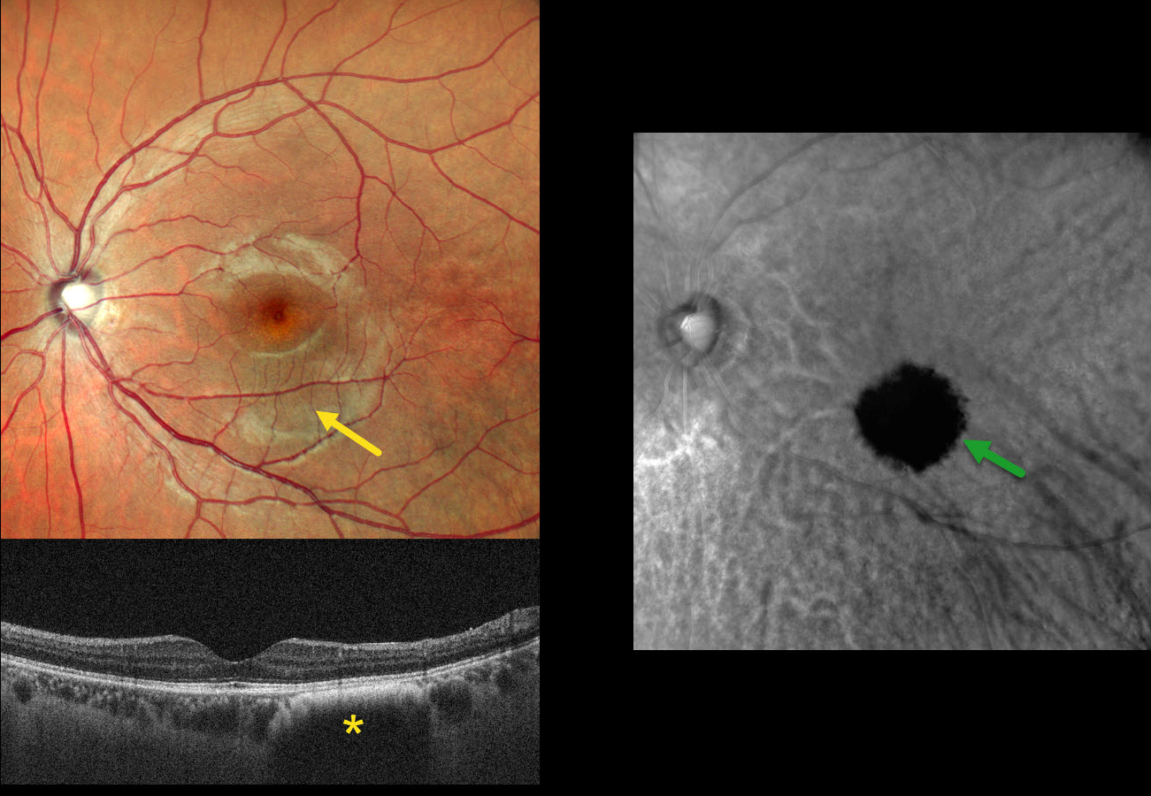
This asymptomatic 26YO male presented for a routine eye examination. Vision was 20/20.
Clinical biomicroscopy and MultiColor scanning laser ophthalmoscope (SLO) imaging are normal except for faint hyperpigmentation in the inferior macula. OCT in this region shows a dome-shaped hyporeflective lesion with an anterior hyperreflective border. Retromode imaging shows a well-defined hyporetroreflective lesion.
Learning Points:
Traditional color photography uses white light to image the posterior segment. Newer imaging devices utilize LED or SLO technology to illuminate the fundus with blue, green, and infrared light, which best image the vitreoretinal interface/inner retina, mid-retina to RPE, and choroid, respectively. Many of these technologies create a ‘color’ fundus image that is not true to life yet contains clinically useful depth information in a flat image. In our patient’s case, Nidek Mirante Retromode imaging best detected this subclinical lesion using a 790 nm SLO. For a recent review of Retromode imaging, see Sukkarieh et al, Surv Ophthalmology 2023;68:1027-1037.
So does our patient have a choroidal nevus or hemangioma? Internally, the authors are split between a nevus (SRS and MN) and a hemangioma (SB). Findings supporting a choroidal hemangioma include its orange color blending with the surrounding RPE/choroid and round shape on Retromode imaging. However, we have not seen a hemangioma with an anterior hyperreflective margin on OCT. Jonna et al described 5 OCT patterns of flat choroidal nevi (Ophthalmology Retina 2019;3:270-277), including a subtype with “anteriorly bowed hyperreflectivity with discrete borders and cascading edges,” which appears identical to our patient’s OCT.



