

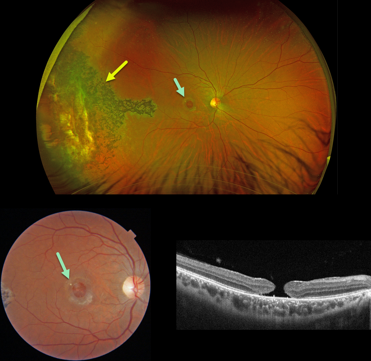
This 31YO male suffered a paint gun injury to his right eye when he was 12YO. Vision has since been stable and poor in this eye. Vision was 20/200.
Optos color RG imaging shows temporal peripheral scarring with intraretinal pigment migration. A large macular hole with surrounding pigmentary changes is noted on Triton color imaging and swept-source OCT.
Learning Points:
Since fresh traumatic holes can spontaneously flatten, especially in pediatric cases (Liu et al, AJO 2020;210:174-183), an initial period of observation is recommended before considering vitrectomy. Given the size and chronicity of our patient’s hole, surgery was not considered.
Photoreceptor loss from numerous causes, including blunt trauma, retinal detachment, macular telangiectasia, and retinitis pigmentosa (RP), can lead to intraretinal RPE migration, a shared phenotype. Unilateral RP is exceedingly rare and requires normal electrophysiologic testing with long-term observation to ensure that delayed findings are present in the fellow eye.



