

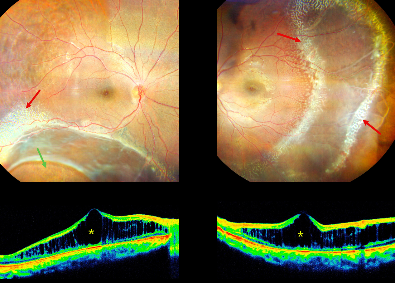
This 16YO male presented with stable bilateral vision loss since childhood, with new floaters in his right eye for 2 weeks. There was no ocular family history. Vision was 20/200 OD and 20/60 OS.
His right eye shows a large area of inferotemporal combined retinoschisis-rhegmatogenous retinal detachment (RD) extending up to the inferotemporal arcade. Subretinal pigmentation is seen along the posterior edge of the schisis inferiorly, and a large oval inner retinal defect is present inferotemporally. A combined schisis-RD is noted in his left eye which extends temporally and inferiorly. Radiating lines of foveal schisis are present bilaterally.
A dendriform pattern of occluded vessels is noted along the superotemporal edge of the schisis OD and surrounding the schisis OS. There are also numerous tiny round inner retinal breaks posterior to these dendriform lesions in the distal temporal left macula. The retinal vessels are variably fibrosed overlying the schisis cavities. OCT scanning shows severe macular schisis, primarily affecting the inner nuclear layer. Bilateral laser demaration was performed along the posterior edges of the schisis-retinal detachments.
Learning Points:
X-linked retinoschisis is caused by a mutation of the RS1 gene which is located on the X chromosome and encodes for retinoschisin, a protein secreted by photoreceptors which is involved in intercellular adhesion and likely retinal cellular organization (Heymann et al, Progress Retinal Eye Research 2023;95:101147). Although usually transmitted as an X-linked recessive disorder in males with an incidence of 1 in 15,000 to 30,000, it can also sometimes affect females.



