

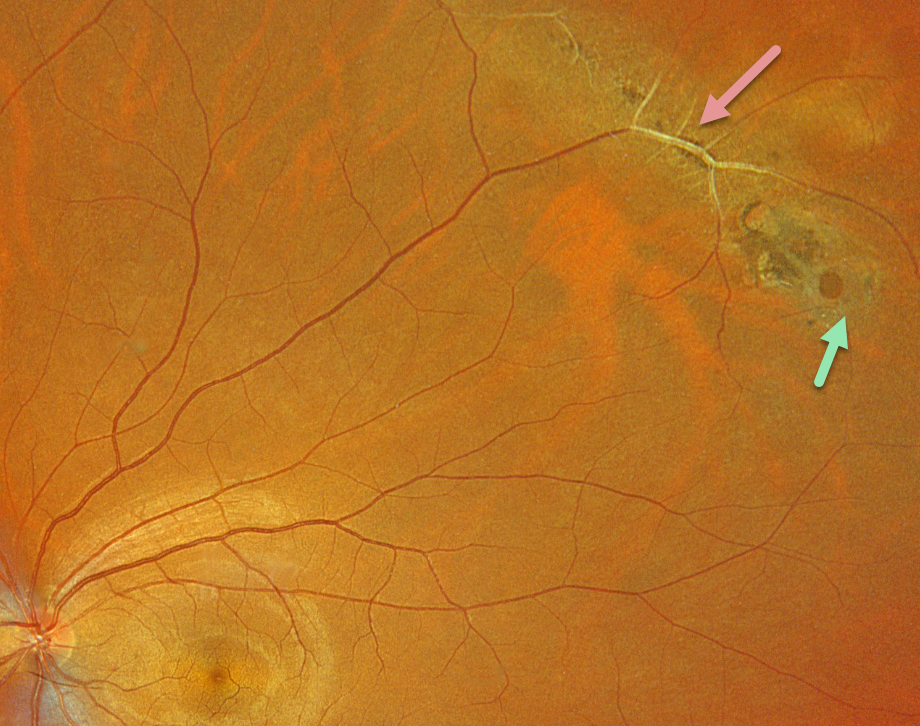
This healthy 33YO male was referred for asymptomatic lattice degeneration in his left eye. Optos color RGB imaging shows a circumferential patch of midperipheral superotemporal paravenous lattice. The large retinal vessels within the lesion are fibrosed. A small atrophic retinal hole is noted along its inferior edge. Observation was recommended.
Learning Points:
Lattice lesions have a pocket of vitreous liquefaction overlying the thinned retina with a firm vitreo-retinal adhesion along their borders. White, fibrosed vessels often crisscross the lesions, hence the name, lattice. This firm vitreoretinal adhesion, along with the often-present atrophic holes, is what can predispose these eyes to retinal detachment.



