

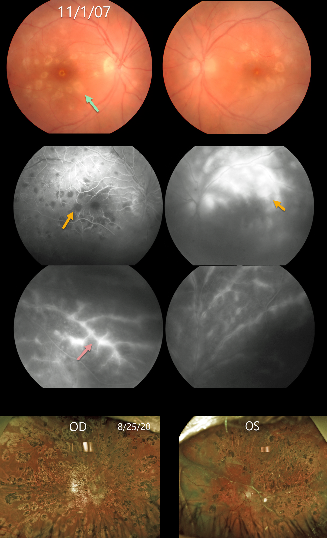
This 18YO female presented on 11/1/07 with acute bilateral panuveitis. Moderate vitreous cells were present along with subretinal multifocal yellow-white choroidal infiltrates. Angiographically, these choroidal lesions blocked early and stained late. A frosted-branch pattern of vascular leakage was noted bilaterally. Following a course of oral prednisone, the acute uveitic findings resolved, leaving behind inactive multifocal scarring. She developed severe recurrent panuveitis a year or two later, leaving her with 20/400 vision bilaterally (images not shown).
Shortly thereafter, she was diagnosed with Hodgkin lymphoma. At almost the exact date of this diagnosis, Baillif et al reported a patient with bilateral angle-closure glaucoma and multifocal choroiditis as the initial presentation for Hodgkin lymphoma (Retinal Cases 2011;5:201-205). The posterior segment findings in this report were virtually identical to those in our patient. The authors felt that the uveitis was a paraneoplastic phenomenon.
She subsequently underwent successful bone marrow transplantation. Her lymphoma and uveitis have both remained in remission since. A rhegmatogenous retinal detachment later developed in her left eye, which was repaired with vitrectomy and silicone oil. Despite successful reattachment, vision subsequently decreased to no light perception. On 8/25/20, vision was 20/400 OD with severe stable bilateral chorioretinal scarring bilaterally.
Learning Points:
Multifocal choroiditis (MFC) is the current preferred term for a group of historically heterogeneous disorders, including multifocal choroiditis, recurrent multifocal choroiditis, multifocal choroiditis and panuveitis, punctate inner choroidopathy, progressive subretinal fibrosis, and pseudo-histoplasmosis (Essex et al, Retina 2013;33:1-4 and Spaide et al, Retina 2013;33:1315-1324). MFC occurs in otherwise healthy adults, often myopic and female. Findings can include variably pigmented multifocal scars throughout the fundus, panuveitis, subretinal fibrosis, and macular neovascularization (MNV). Unlike ocular histoplasmosis, new scars and uveitis can be seen. MNV can be successfully managed with anti-VEGF therapy.
As with any uveitis, the clinician must always include the possibility of a masquerade syndrome as occurred in our patient.



