

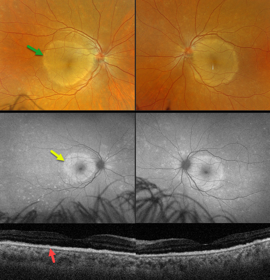
This 45YO female presented with one week of bilateral vision loss. Vision was counting fingers in each eye.
Optos color RGB imaging shows symmetrical, discrete subretinal yellow-white placoid lesions occupying each macula. Fundus autofluorescence (FAF), however, shows multifocal hyper-FAF spots extending peripherally to the hyper-FAF macular lesions. Swept-source OCT shows replacement of the outer retinal bands with hyperreflective material.
Laboratory testing was positive for syphilis and negative for HIV. She was referred to an infectious disease specialist for systemic penicillin treatment. Unfortunately, she was immediately lost to follow-up.
Learning Points:
Ocular manifestations of syphilis vary widely and include virtually all variations of anterior and posterior segment inflammation. Originally described by Gass (Ophthalmology 1990;97:1288-1297), placoid chorioretinitis is one of the more common presentations. The discrete placoid lesions were quite dramatic on our patient’s multimodal imaging.



