

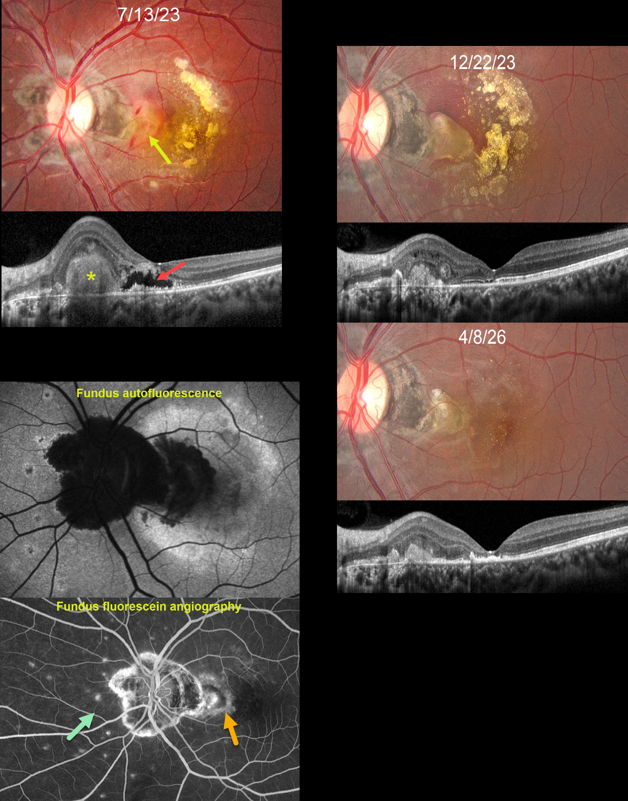
This 21YO female presented with a gradually progressive central scotoma in her left eye for the prior 2 months. Vision was 20/20 in her normal OD and 20/1600 OS.
Fundus photography shows a foveal subretinal fibrotic macular neovascularization (MNV) with surrounding subretinal blood and variably confluent subretinal lipid noted more temporally. Some inner retinal hemorrhages are also noted. Variably pigmented peripapillary scarring is present with multifocal small, depigmented scars nasally. OCT scanning shows a hyperreflective subretinal mound corresponding to the area of subretinal fibrosis. More centrally, there is a fluid-filled pocket of outer retinal hyporeflectivity surrounded by a ragged, variably thickened border of hyperreflective material. Fundus autofluorescence (FAF) shows peripapillary hypo-FAF as does the subretinal fibrosis with surrounding blood. The macula otherwise shows variable hyper-FAF, likely due to unmasking from outer retinal atrophy. The MNV stains angiographically. Multifocal scars stain superiorly, nasally, and inferiorly.
Following 3 monthly injections of Eylea, vision improved to 20/50. The MNV is contracted into an oval-shaped subretinal lesion with surrounding fading subretinal blood and absorbing lipid. She was then lost to follow-up for over 2 years. Thankfully, the MNV showed further contraction, with faint, scattered residual lipid dots. Despite outer retinal atrophy on OCT, vision improved to 20/25.
Learning Points:
Multifocal choroiditis (MFC) is the current preferred term for a group of historically heterogeneous disorders, including multifocal choroiditis, recurrent multifocal choroiditis, multifocal choroiditis and panuveitis, punctate inner choroidopathy, progressive subretinal fibrosis, and pseudo-histoplasmosis (Essex et al, Retina 2013;33:1-4 and Spaide et al, Retina 2013;33:1315-1324).
MFC occurs in otherwise healthy adults, often myopic and female. Findings include variably pigmented multifocal scars throughout the fundus, panuveitis, subretinal fibrosis, and MNV. Unlike ocular histoplasmosis, new scars and uveitis can be seen. MNV can be successfully managed with anti-VEGF therapy.
Our patients’ MNV and peripheral scars most likely represent MFC. We continue to follow her closely



