

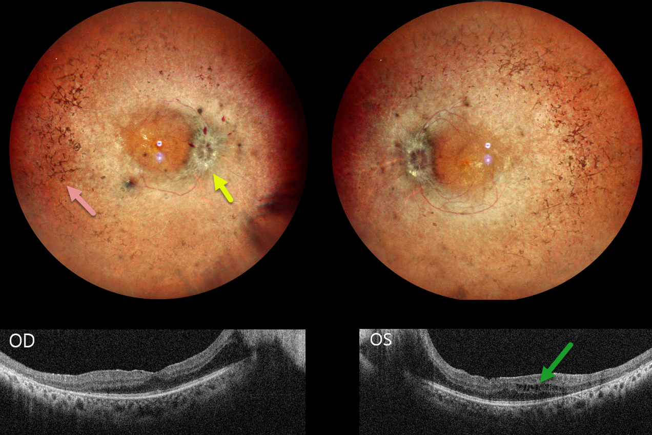
This 30YO male presented with 3 weeks of decreased vision. His father has a history of retinitis pigmentosa (RP). Vision was 20/30 OD and 20/80 OS.
Pseudocolor SLO imaging shows typical bilateral RP scarring with intraretinal pigment migration extending from the peripheral maculas into the retinal peripheries. Bilateral macular puckers and disc swelling with right peripapillary inner retinal fluid are also seen. OCT scanning confirms the epimacular membranes. Bilateral pericentral outer macular atrophy is noted, along with some cystic edema in the left inner and outer nuclear layers.
Learning Points:
RP is a clinically and genetically heterogeneous group of inherited retinal disorders. The disease can present sporadically or with any inheritance pattern (autosomal dominant, autosomal recessive, sex-linked, or mitochondrial). It is characterized by diffuse, progressive dysfunction of predominantly rod photoreceptors, with subsequent RPE degeneration and intraretinal pigment migration. Visual impairment usually presents as night blindness and progressive visual field loss.
Bilateral optic disc edema is not typical for RP. Unfortunately, we were unable to work up our patient for a secondary cause because he was lost to follow-up immediately.



