

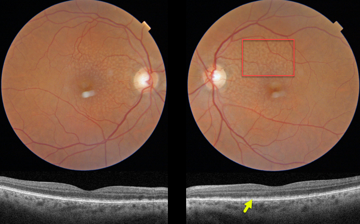
This 70YO female was referred for asymptomatic age-related macular degeneration. Vision was 20/30 OD and 20/25 OS.
Triton color imaging of both eyes shows symmetrical ribbon subretinal drusenoid deposits. On swept-source OCT, these lesions appear as hyperreflective material mostly between the RPE and ellipsoid zone (EZ).
Learning Points:
Subretinal drusenoid deposits (SDD) are polymorphous light-gray interconnected accumulations located above the RPE. They are present in about one-quarter of older adults with healthy maculas and in more than half of those with early-to-intermediate dry AMD. They can appear as an array of dots, interconnected reticular bands (as in our patient), or confluent lesions (Suzuki and Spaide, AJO 2014;157:1005-1012). The dot variant is associated with macular neovascularization (MNV) and the confluent variant with macular atrophy (Zhou et al, Ophthalmology 2016;123:1530-1540).
Clinically, they are often mistaken for conventional drusen and consist of similar yet distinct lipid-rich material found in soft drusen. They are localized to rods, unlike typical soft drusen, which are more central and cone-based. As with drusen, SDD are dynamic and can grow, shrink, resolve, or even recur.
Since they are located above the RPE, they are best visualized with blue or green illumination. On OCT, they appear in 3 stages (Zweifel et al, Ophthalmology 2010;117:303-312) growing upwards from the RPE towards the external limiting membrane: stage 1, diffuse deposition of granular hyperreflective material between the RPE and the EZ, stage 2 material alters the EZ contour, and stage 3 conical appearance breaking through the EZ. Rod function seems to be preferentially affected, and there is an associated increased risk of delayed dark adaptation, macular atrophy and types 2 and 3 MNV. In fact, type 2 neovascularization in the setting of AMD occurs almost solely in eyes with subretinal drusenoid deposits.
In addition to large drusen and pigmentary abnormalities, SDD is the 3rd risk factor for progression to late AMD (Agron et al, Ophthalmology 2022;129:1107-1119). For a recent review, see Wu et al, Progress Retinal Eye Research 2022;88;101017.



