

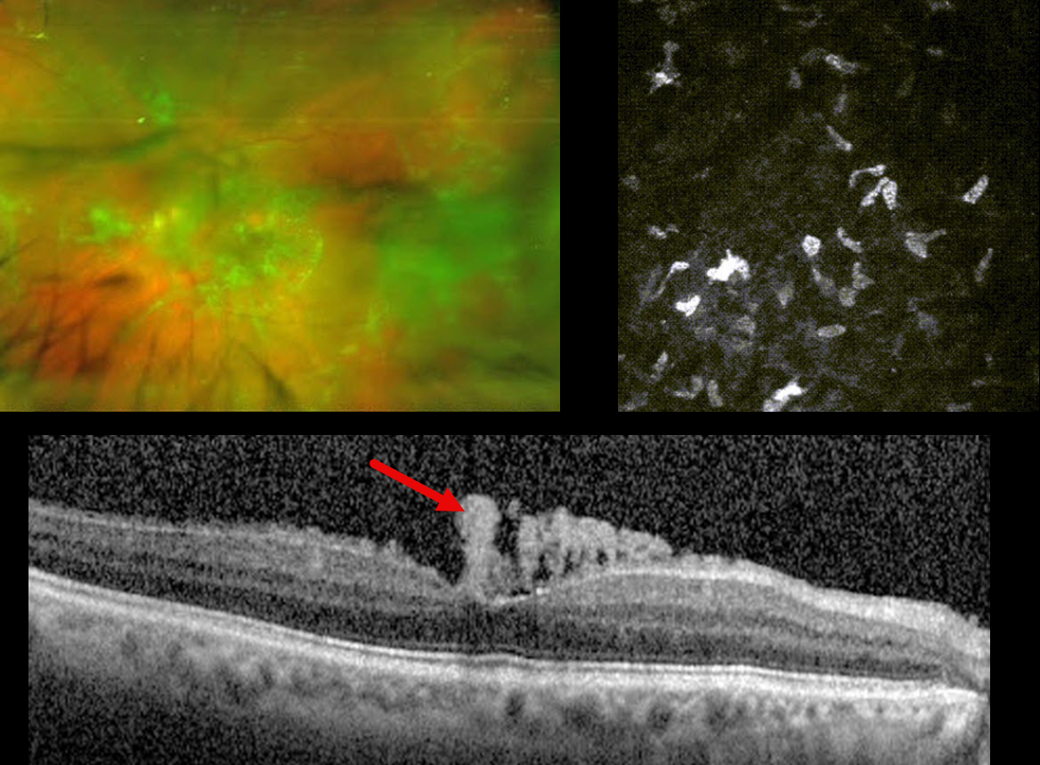
This 65YO male presented with chronic bilateral vision loss. Vision was 20/200 OD and 20/100 OS. There was no past medical history.
Optos color RG imaging of his right eye shows diffuse whitish, spider web-like fibrillar vitreous debris. In vivo corneal confocal microscopy reveals amyloid deposits.
Sequential therapeutic and diagnostic vitrectomy surgeries were performed on each eye. Vitreous and conjunctival biopsies were positive for amyloid deposits. Each fundus appeared normal postoperatively, and vision improved to 20/20 OU. Despite the normal fundus appearance, OCT scanning shows residual vertical amyloid deposits resting on the foveal internal limiting membrane.
Similar findings were present in his left eye (not shown).
Learning Points:
Vitreous amyloidosis is a rare disorder where bilateral vitreous opacities usually appear as fine, fibrillar, and often whitish or translucent deposits, which can reduce vision as the opaque amyloid material accumulates. OCT can reveal pathognomonic fine vertical needle-like hyperreflective deposits extending into the vitreous from the internal limiting membrane (Tasiopoulou et al, Ophthalmology Retina 2021;5:99-101). It is almost always related to mutant transthyretin (TTR), a plasma protein that transports thyroxine and vitamin A, often associated with familial amyloid polyneuropathy (Venkatesh et al, Ophthalmology 2017;124:1014-1022). Systemic amyloidosis can also cause ocular manifestations, including conjunctival, temporal artery, extraocular muscle, trabecular meshwork, and cranial nerve deposition (Reynolds et al, Retina 2018;38:1371-1376).
Our patient’s systemic workup was negative. Genetic testing for TTR was ordered and is pending.



