

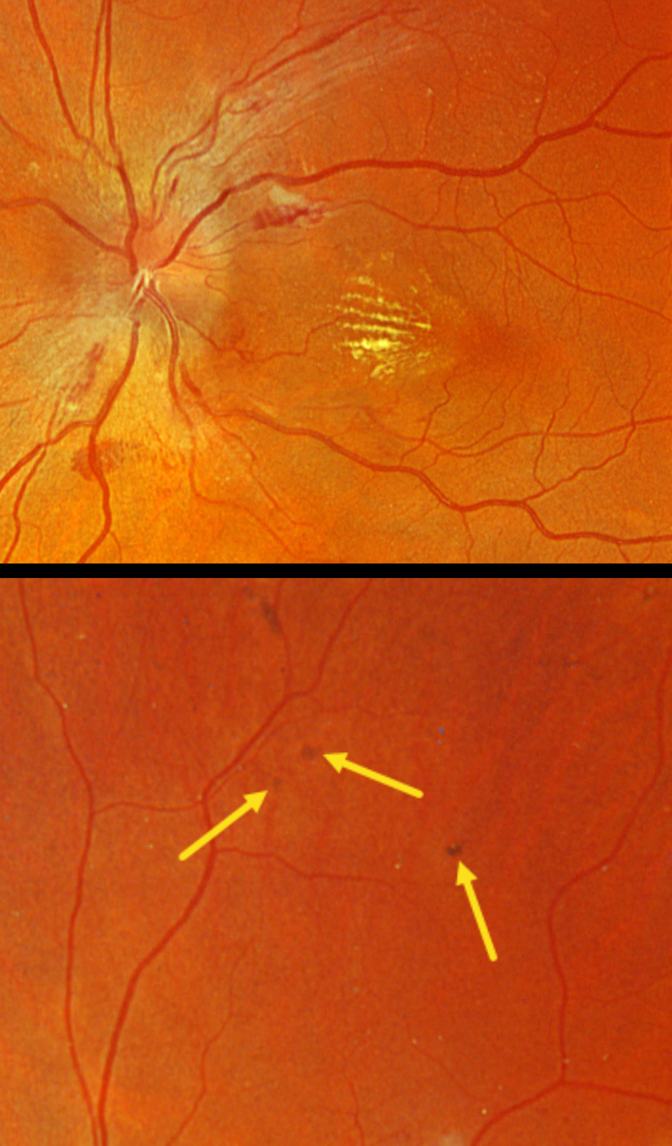
This 46YO male presented to his local optometrist 1 week earlier with a few days of bilateral vision loss. His doctor diagnosed malignant hypertension due to retinal findings and a blood pressure of about 280/160. He was hospitalized for a few days and presented to us for further evaluation. Vision was 20/30 OD and 20/50 OS.
Optos color RGB imaging shows optic nerve edema, peripapillary hemorrhages, and a nasal macular lipid star. Several small pigmented Elschnig spots are noted superiorly OS. He was immediately lost to follow-up.
Learning Points:
Malignant hypertension, defined as blood pressure above 180 systolic and/or 120 diastolic, is a life-threatening medical emergency. Eye doctors are in a unique position to often diagnose this condition. Patients will often present with bilateral optic nerve swelling, flame-shaped retinal hemorrhages, and, with more chronic disease, lipid precipitates in the nasal macular outer plexiform layer. Focal (Elschnig spots) or linear (Siegrist streaks) choroidal pigmentary changes develop from hypertensive choroidopathy.



