

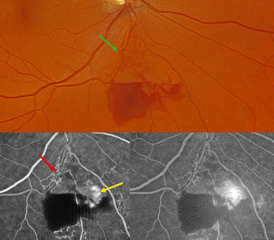
This 59YO male was referred for asymptomatic retinal findings in his right eye. Vision was 20/30.
Optos color RG imaging shows an inferior branch retinal vein occlusion with midperipheral preretinal blood. Collateral vessels are noted at the site of the occlusion. These collaterals show no leakage angiographically, although the adjacent retinal neovascularization does leak.
Learning Points:
Collateral vessels are dilated, pre-existing vessels that bypass an occlusion. These are most commonly seen in the posterior pole following retinal venous occlusions and are exceedingly rare with arterial occlusions. The endothelial tight junctions are intact, so they don’t leak angiographically. This helps differentiate them from neovascularization, which does leak. Our patient’s eye shows us a wonderful example of both.



