

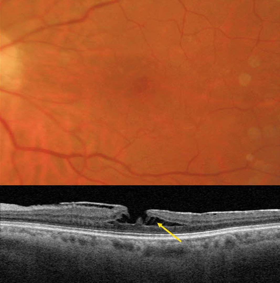
This 62YO female has a known history of an asymptomatic macular pucker. Vision was 20/70.
Triton color imaging shows a faint, irregular macular pseudohole. Swept-source OCT shows a variably adherent hyperreflective macular pucker. A tractional lamellar macular hole (LMH) is noted centrally, with pillars of Müller cells spanning the split outer plexiform layer (OPL).
Learning Points:
LMH can be defined as tractional or degenerative (Govetto et al, AJO 2016;164:99-109). Tractional LMH shows OPL splitting, an intact ellipsoid zone, and is associated with vitreomacular traction and tractional epiretinal membranes. OCT features of a degenerative LMH include loss of outer retinal tissue below the OPL and an outer retinal ‘bump’. Non-tractional epimacular proliferation is often present and will often extend around the posterior edge of the degenerative LMH.



