

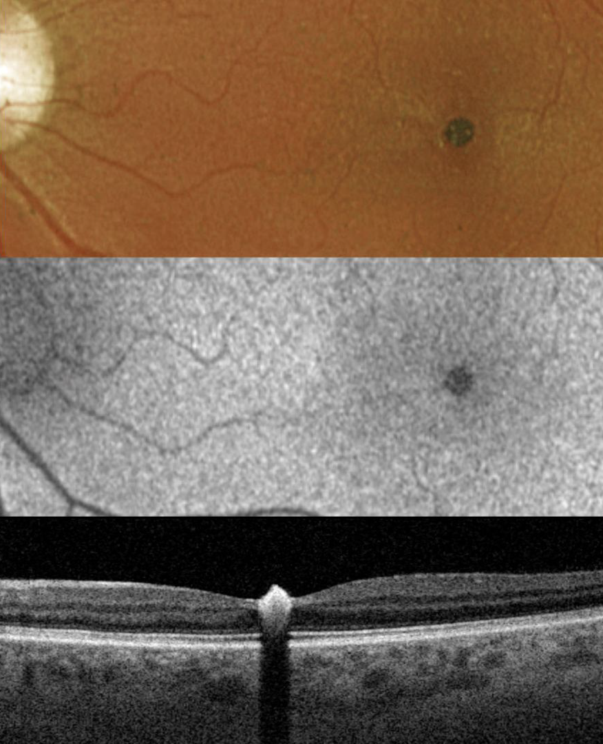
This 38YO male was referred for an asymptomatic lesion in his left eye. Vision was 20/25 in his normal OD and 20/25 OS.
Optos color RG imaging shows a small foveal hyperpigmented lesion that hypo-autofluoresces. Triton swept-source OCT shows a markedly hyperreflective intraretinal lesion that protrudes above the retinal surface with marked posterior shadowing. Observation was recommended.
Learning Points:
RPE hamartoma is an extremely rare, benign, presumed congenital lesion. It appears as a small, jet-black inner vs full-thickness foveal or juxta-foveal retinal lesion (Shields et al, Ophthalmology 2003;110:1005-1011). These lesions are markedly hyperreflective on OCT (Takahashi et al, Retinal Cases & Brief Reports 2013;7:179-182).



