

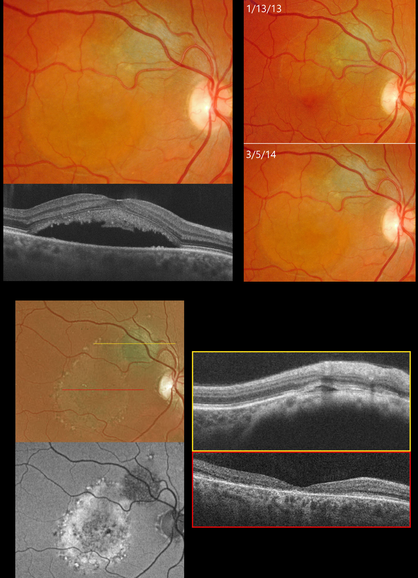
This 35YO female was examined for a suspect choroidal nevus on 1/13/13. Color photography shows about a 2x2mm minimally thickened choroidal nevus superotemporal to the right optic nerve (image 2). Subretinal fluid extends into the superior macular and some orange pigment overlies the nevus.
She returned on 3/15/14, now with fluid extending through the fovea (image 1). OCT scanning confirms the foveal fluid and hyperreflective material, likely representing shed outer segments, lines the outer surface of the detached macula.
She was referred to an ocular oncologist and immediately lost to follow-up until she presented on 4/2/25 without new complaints. She told us she had received two photodynamic therapies elsewhere shortly after we last saw her over 10 years earlier. Vision was 20/200. Optos color RG imaging shows the nevus to be stable in size but with new pigmentary changes throughout the macula within the area of prior fluid (image 3). On fundus autofluorescence (FAF), the nevus is hypo-FAF, and the region of prior fluid shows variable hyper-FAF. OCT scanning shows no fluid overlying the nevus with foveal atrophy. Continued observation was recommended.
Learning Points:
Clinical characteristics of choroidal nevi at risk for progressing to a uveal MM were initially described by Shields et al (Ophthalmology 1995;102:1351-1361). The most recent version of this famous acronym (Shields et al, Retina 2019;39:1840-1851) is To Find Small Ocular Melanoma Doing Imaging (TFSOM-DIM), which stands for To (Thickness >2mm) Find (subretinal Fluid) Small (Symptoms, vision <= 20/50) Ocular (Orange pigment) Melanoma (Melanoma hollow on ultrasonography) DIM (DIaMeter > 5mm).



