

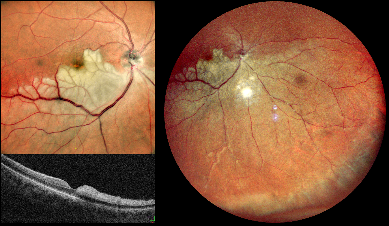
This is a healthy 42YO male presented with 7 days of blurred vision in his left eye. Vision was 20/90 OD and 20/20 in his normal OS.
Pseudocolor SLO imaging shows a white, opaque retina in the inferior retinal hemisphere, which spares the central macula. The vertical B-scan contrasts the normal superior retina with the ischemic inferior retina with marked inner retinal hyperreflectivity.
Learning Points:
Once the central retinal artery passes anteriorly through the lamina cribrosa, it branches into superior and inferior branches, which then further subdivide to supply the inner two-thirds of the neurosensory retina. Hemi-CRAO is a rare CRAO variant caused by the blockage of one of these superior or inferior branches. As with more typical CRAO, emboli or giant cell arteritis are the most common etiologies.
Our patient had acute retinal ischemia (defined as acute BRAO, central retinal artery occlusion, or transient monocular vision loss) and was immediately referred to the nearest stroke center. Unfortunately, he was immediately lost to follow up.



