

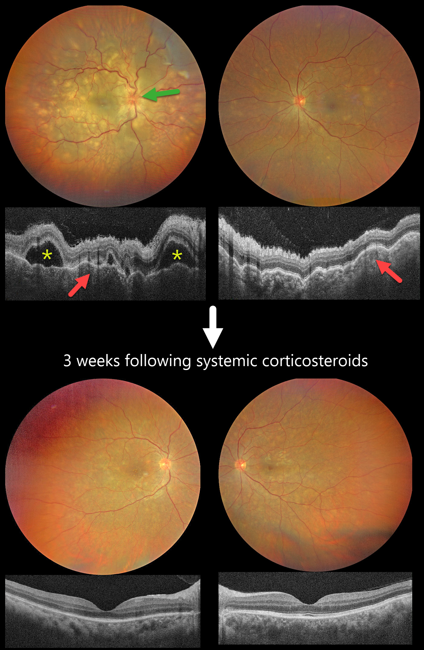
This previously healthy 40YO female presented with 2 days of blurred vision. She denied any systemic symptoms. Vision was 20/200 OD and 20/60 OS.
Color photography shows multifocal yellow-white subretinal lesions scattered throughout each posterior pole, with the right eye greater than the left, associated with overlying multifocal exudative retinal detachments. The right nerve is also edematous. OCT scanning shows a bilateral thickened choroid with overlying chorioretinal folds and multifocal serous detachments OD.
Intravenous methylprednisolone (1000mg/day) was administered for five days, followed by a 2-week course of oral prednisone (1mg/kg). Three weeks later, the funduscopic and OCT findings normalized. Vision improved to 20/30 OD and 20/20 OS.
Learning Points:
Harada disease is characterized by relatively acute bilateral panuveitis, optic nerve swelling, thickened choroid, chorioretinal folds, multifocal serous exudative retinal detachments, and bacillary layer detachments. Patients with just ocular findings have Harada disease (our patient), whereas those with additional systemic findings (including vitiligo, poliosis, headache, vertigo, and hearing loss) have Vogt-Koyanagi-Harada disease.



