

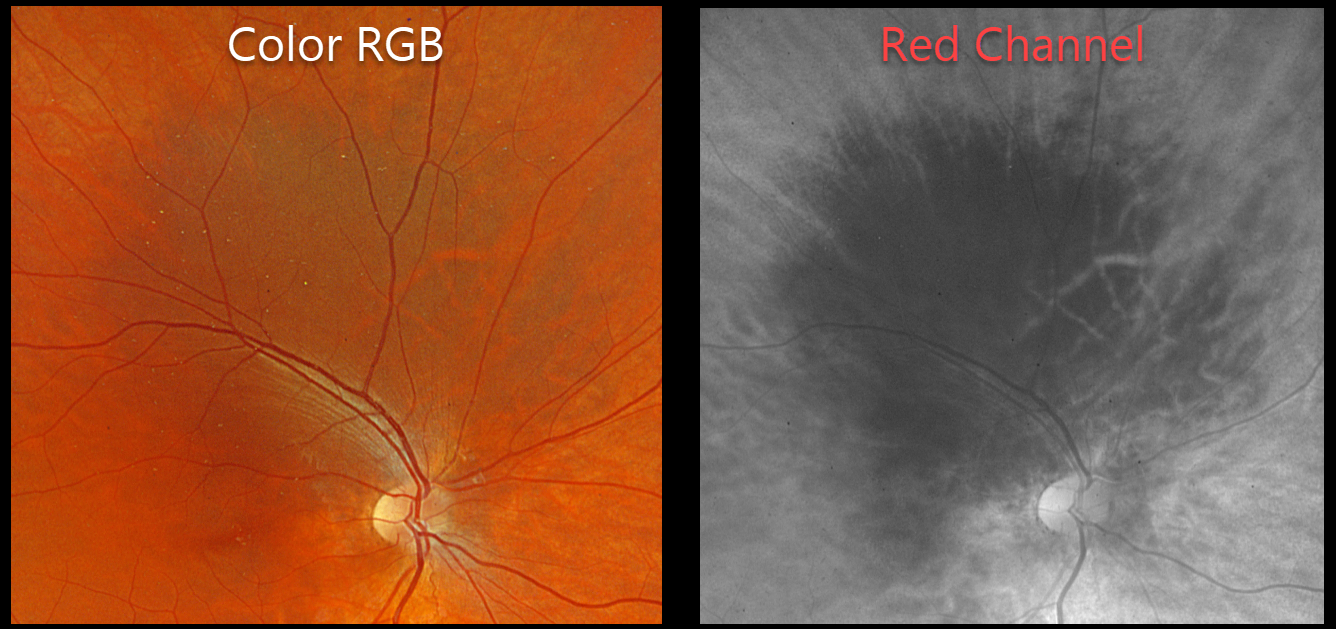
This healthy 15YO male was referred for an asymptomatic pigmented lesion in his right eye. Family history was negative. Vision was 20/40 OD and 20/30 in his normal OS.
Optos color RGB imaging shows a region of increased choroidal pigmentation extending from the superonasal macula into the superior midperiphery. This lesion is best seen with the red channel.
Learning Points:
Choroidal melanocytosis is part of the oculodermal spectrum which occasionally includes pigmentation of the globe or periocular skin (Nevus of Ota, melanosis oculi). There are increased dendritic melanocytes in the affected tissues following the distribution of the first and second branches of the trigeminal nerve. Patients need ongoing monitoring since about 1 in 400 will develop uveal melanoma. Of those that develop melanoma, they are twice as likely to undergo metastasis when compared to patients with uveal melanoma and no pre-existing melanocytosis.
Although pretty color images that match what we see on biomicroscopy are most intuitive clinically, newer imaging modalities that distort the normal fundus colors often contain very clinically relevant information. Standard Optos color RG images are generated from a red (635nm) and green (532nm) laser. Although these images have a greenish tint, they essentially contain two images in one: the RPE/neurosensory retina is best captured with the green channel, and the choroid is best seen with the red channel. The newer Optos true color RGB unit maintains the ability to view the separate RG channels but also generates a true to life color image by adding a blue (488nm) laser. In our patient’s case, the red channel from the RG image best shows the melanocytosis.



