

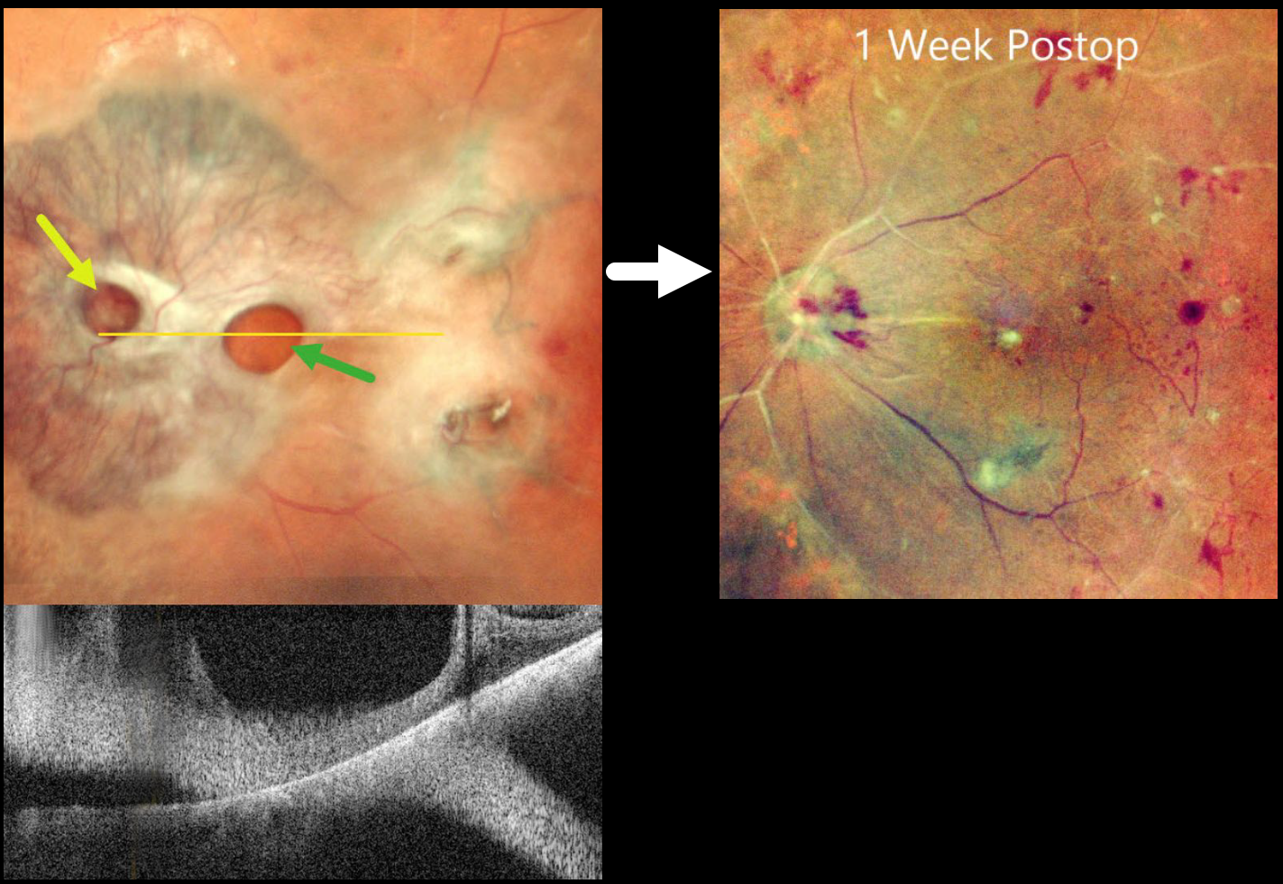
This 65YO diabetic female presented with counting fingers bilaterally from proliferative diabetic retinopathy with macular tractional retinal detachments.
Preoperative pseudocolor SLO imaging of her left eye shows a complex fibrovascular membrane covering the posterior pole. Pseudo giant-macular holes are noted overlying the disc and macula. OCT scanning confirms the lack of a true macular hole. One week following vitrectomy, membrane peeling, and air injection, vision improved to 20/200, and the retina was completely attached with no residual traction.
Learning Points:
A giant macular hole, also reported in Alport syndrome (Shah and Weinberg Ophthalmic Genetics 2010;31(2):93-97), is an exceedingly rare traumatic event (Hernandez-Da Mota Case Rep Ophthalmol 2011;2:283-286). The unusual anatomy of our patient’s fibrovascular diabetic membrane simulated a giant macular hole.



