

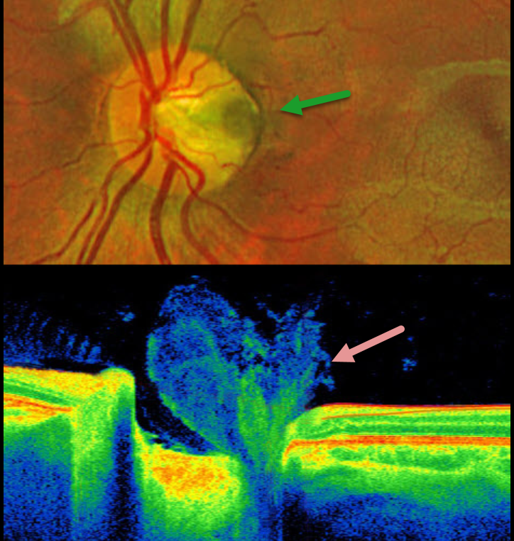
This healthy 6YO boy presented for a routine examination. Vision was 20/20 in his normal right eye and 20/20 in his left eye. OCT scanning shows a vitreous plug extending into the depth of the pit.
Optos color RG imaging shows a temporal optic nerve pit without associated macular abnormalities.
Learning Points:
An optic nerve pit is a rare (about 0.1% prevalence) congenital anomaly thought to be caused by incomplete closure of the optic fissure during gestation. Pits are most often located within or along the inferior-temporal disc margin and usually appear as a gray depression. While most optic pits are asymptomatic, they can cause vision loss due to fluid being forced into the inner retina, outer retina, and the subretinal space (optic pit maculopathy).
Utilizing high-resolution swept-source OCT, Spaide defined the optic pit complex as a broader means to characterize the structural abnormalities found with optic disc pits (Retina 2026;46:15-24). The spectrum of abnormalities includes lamina cribrosa defects (all eyes) with associated tissue cavitations extending a mean of 1855 microns posterior to a modified Bruch membrane opening. One of his patients had OCT findings similar to our patient’s, with vitreous extending into the pit. Sources of intra- and subretinal fluid included direct communication from a cavitation, perivascular hyporeflective spaces, intraneural channels extending toward cystoid spaces, and isolated retinal cysts. These concepts support the “dynamic and multifactorial process involving abnormal structural anatomy, disrupted tissue boundaries, potential misrouting of glymphatic flow, vitreous or cerebral spinal fluid (CSF)-related gradients, and mechanical forces exerted during eye movements.”



