

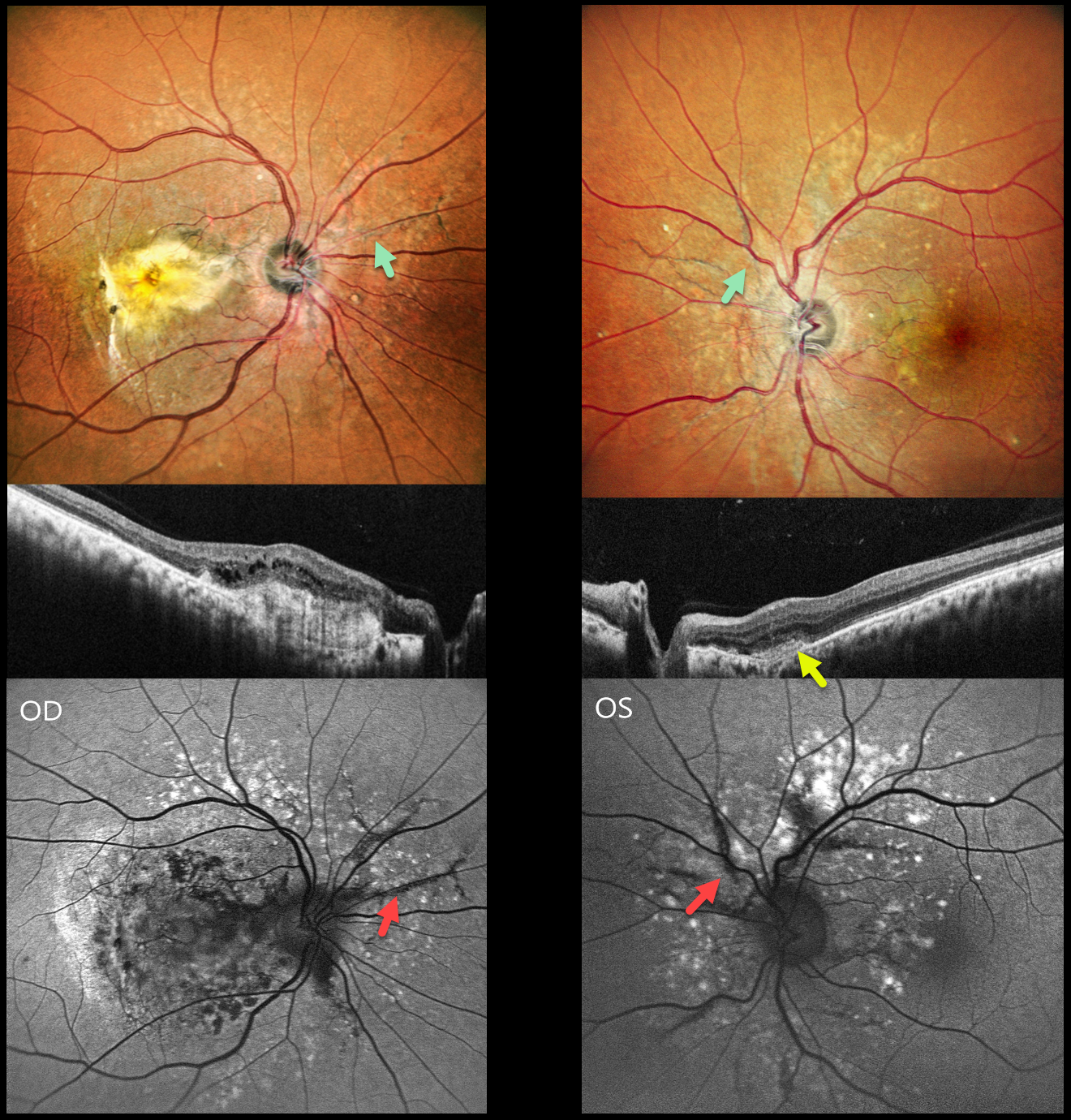
This healthy 42YO male presented with 10 months of vision loss in his right eye and 1 month of mild blurred vision in his left eye. Vision was counting fingers OD and 20/20 OS.
Pseudocolor SLO imaging shows angioid streaks extending around and radiating outwards from each optic nerve. There are drusen-like changes around each posterior pole, along with clumps of subretinal pigment temporally.
OCT scanning shows hyperreflective subretinal fibrosis with mild overlying cystic retinal edema OD. A much smaller area of subretinal hyperreflectivity (indicating a type 2 macular neovascularization, MNV) with overlying subretinal hyperreflective material is noted OS. The angioid streaks on fundus autofluorescence (FAF) are hypo-FAF due to RPE atrophy. The drusen-like changes are hyper-FAF.
Learning Points:
Retinal findings in pseudoxanthoma elasticum (PXE) most commonly include angioid streaks and a subretinal peaux d’orange appearance, most pronounced in each temporal macula. However, pattern dystrophy-like changes, as in our patient, can sometimes be the predominant phenotype (Murro et al, Graefe’s 2020;258:1881-1892). As with typical pattern dystrophies, the findings are dynamic and best captured with multimodal imaging.
PXE is caused by a mutation in the ABCA6 protein and is virtually always transmitted as an autosomal recessive disorder, although autosomal dominant transmission may rarely occur (Plomp et al, Am J Med Genet A 2004;126:403-412).
Our patient had no clinical signs of PXE and denied any family history of this disease. He was referred to his primary care doctor for further evaluation. Intravitreal anti-VEGF therapy was started OS.



