

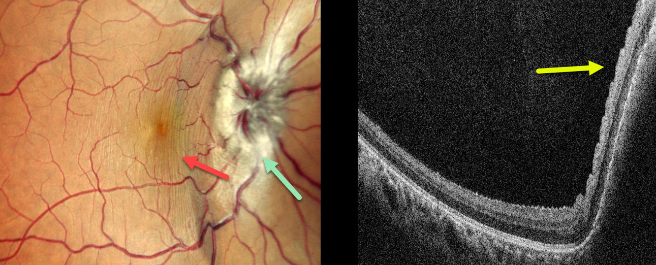
A 26YO male presented with 3 weeks of right eye pain, proptosis, and vision loss. MRI several days earlier was suggestive of an orbital venolymphatic malformation (VLM). Vision was 20/30 OD and 20/20 in his normal OS. The right eye was grossly proptotic.
Multicolor SLO imaging shows right disc edema with nasal macular linear inner retinal folds circumferential to the nerve. On OCT, these folds appear as corrugations along the nasal macular surface.
Learning Points:
Orbital and periorbital VLMs are benign congenital vascular lesions that represent 1-3% of all orbital masses. These are composed of abnormal vascular channels lined by endothelial cells, with a spectrum ranging from venous to lymphatic characteristics. Clinically, these present with progressive ptosis, proptosis, restriction of eye movements, swelling, and, in severe cases, with acute visual loss due to intralesional hemorrhage or optic nerve compression. Treatment includes surgery for superficial VLM and intralesional sclerotherapy with bleomycin for non-resectable or treatment-resistant tumors. Ultrasound-guided intralesional bleomycin was recommended for our patient.
In 1911, Paton and Holmes established that papilledema was due to elevated intracranial pressure rather than inflammation, as was previously thought (Brain 1911;33:389-432). They also noted that nerve edema was associated with “a series of folds which run concentric with the edge of the disc,” which they felt represented retinal folds. Sibony and Kupersmith, correlating OCT with fundus photography, found that these folds represented a variety of structural stresses on the nerve and peripapillary tissues, including peripapillary wrinkles, inner retinal folds, choroidal folds, and outer retinal folds/creases (Ophthalmology 2016;123:1397-1399).



