

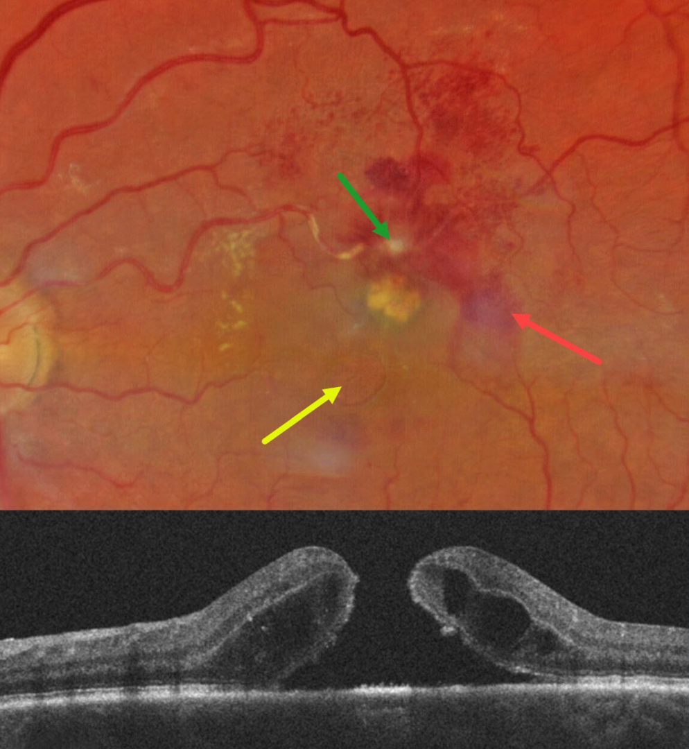
This 67YO white female presented for her six-month diabetic follow-up with a few weeks of vision loss in her left eye. Vision was 20/80 OS and 20/20 in her normal OD.
Color photography shows a fibrosed retinal arterial macroaneurysm (RAM), in the superior macula. Faint subretinal blood is noted temporally, with mostly inner retinal blood superotemporally. The artery is sheathed just proximal to the RAM. A large macular hole with surrounding subretinal fluid is present, accompanied by a lipid clump between the hole and the RAM. Faint lipid is also noted in the nasal macula. She was referred to a retinal specialist for further management.
Learning Points:
Macular hole is a rare finding with retinal arterial macroaneurysms (Sagara et al, Br J Ophthalmol 2009;93:1337-1340). Subretinal and sub-internal limiting membrane blood appears to increase the risk of macular hole.



