

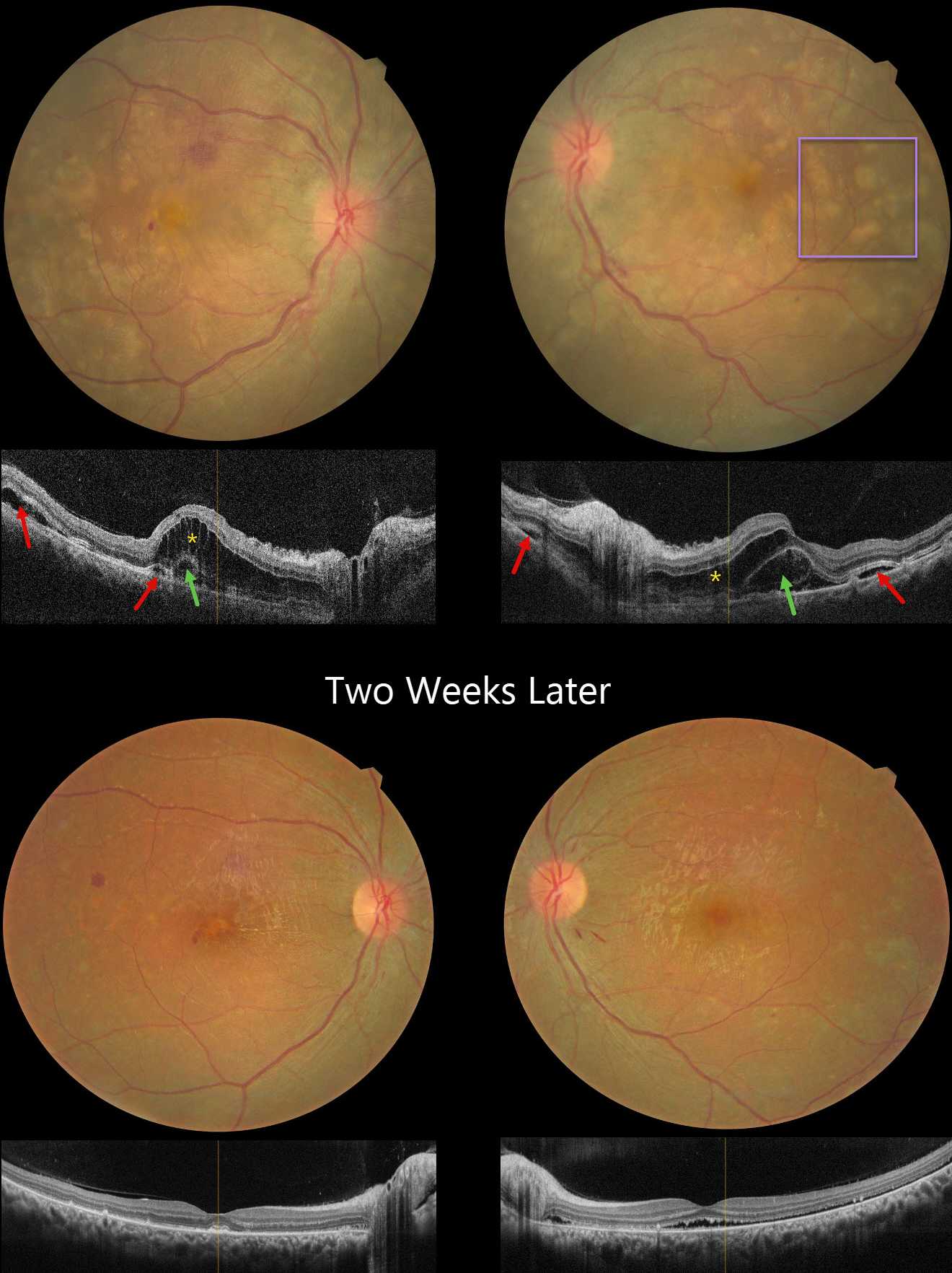
This 24YO female presented with several days of bilateral vision loss. She has a history of anti-glomerular basement membrane disease (anti-GBM) and is on hemodialysis. Her renal disease was quiescent, and there were no acute systemic complaints. There was no anterior segment inflammation, although mild vitreous cells were present bilaterally. Vision was 20/200 OD and 20/80 OS.
Fundus photography shows bilateral multifocal yellow subretinal lesions, inner retinal folds, some retinal hemorrhages, and mild disc edema. OCT scanning shows pockets of subretinal fluid, outer nuclear layer edema, and bacillary layer detachments. The choroid was thickened in both eyes on ultrasonography (not shown).
Oral prednisone 60mg/day was started in a tapering dose. Two weeks later, vision improved to 20/40 OD and 20/80 OS. The acute fundus findings were mostly resolved bilaterally.
Learning Points:
Harada disease is characterized by relatively acute bilateral panuveitis, optic nerve swelling, thickened choroid, chorioretinal folds, multifocal serous exudative retinal detachments, and bacillary layer detachments. Patients with just ocular findings have Harada disease, whereas those with additional systemic findings (including vitiligo, poliosis, headache, vertigo, and hearing loss) have Vogt-Koyanagi-Harada disease.
Anti-GBM disease is a rare autoimmune disorder characterized by autoantibodies that primarily target the basement membranes of the capillaries in the kidneys and lungs. Retinal findings include drusen occurring at an early age (Mansour et al, AJO Case Reports 2017;7:83-90). Acute fundus findings are rarely reported and consist of possibly steroid-induced central serous-like changes with multifocal retinal pigment epithelial detachments and exudative retinal detachments.



