

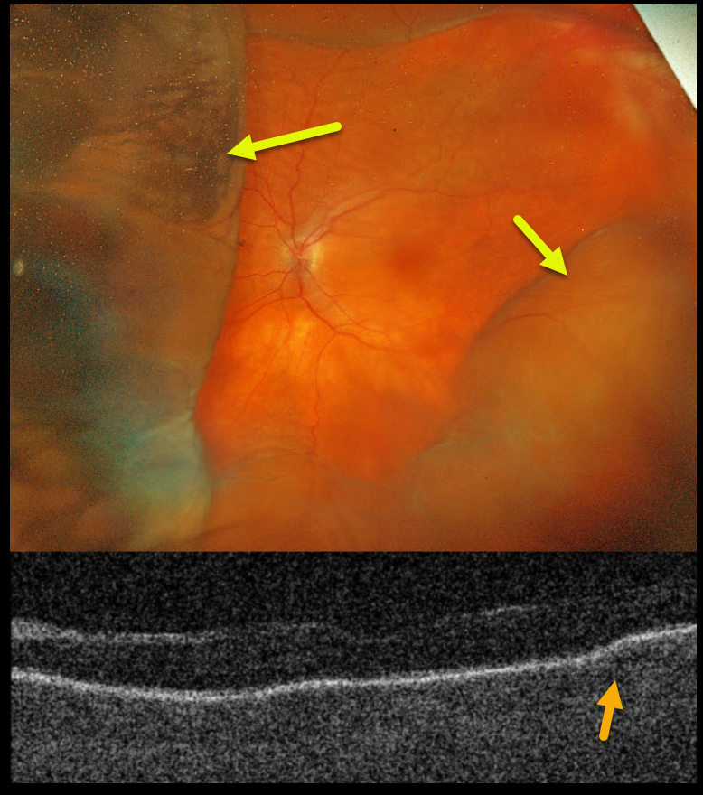
This 78YO female presented with acute vision loss in her left eye. Penetrating keratoplasty (PKP) was performed 9 months earlier. When seen on 8/26/24, vision was counting fingers in part due to irregular astigmatism from the residual PKP sutures, which were then removed. The cornea was Seidel negative after removal. She returned one week later complaining of a curtain in her visual field. Her anterior chamber was flat, and her corneal graft was dehisced for 3 clock hours.
Optos color RGB imaging shows a variably elevated choroidal detachment. Triton swept-source OCT shows chorioretinal folds.
Learning Points:
A choroidal detachment is defined by the abnormal presence of fluid or blood in the suprachoroidal space. Serous choroidal detachments, also known as choroidal effusions, are a frequent complication of glaucoma surgery. Several causative mechanisms are believed to be at play: hypotony allows fluid to accumulate in the suprachoroidal space and inflammation increases the choroidal permeability. The detached ciliary body may also produce less aqueous, thus creating a self-perpetuating cycle. Often, observation is sufficient but topical steroids and cycloplegics can be helpful to aid in resolution.
Hypotony maculopathy is characterized by low IOP and chorioretinal folds, most commonly following glaucoma filtration surgery. Optic nerve edema can also be seen due to decreased axoplasmic transport.
Our patient was immediately taken to surgery for suturing of the dehisced PKP. The choroidals and chorioretinal folds immediately improved and completely resolved two months later (not shown).



